Coronary Angiography
Coronary angiography read: vessels, SYNTAX segments and stenosis with QCA evidence.
Cardiac13 sections
XA· cinefrom $0.12 / request
IVOCT pullback read: lumen, plaque phenotype, calcium, stent sizing and strut-level stent analysis.
This model is not available to run yet. Vote to show interest and get an email when it opens.
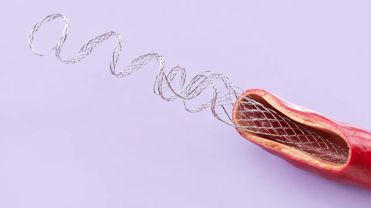
Intravascular OCT reads one coronary optical coherence tomography pullback. At an axial resolution of 10–20 micrometres, OCT shows fibrous-cap thickness, calcium thickness and single stent struts, but analysing them frame by frame is labour-intensive and mostly left to core labs.
The run traces the lumen on every frame, classifies the plaque along the pullback with lipid arc and fibrous-cap thickness, measures calcium arc, thickness and length with the OCT calcium score, proposes stent-sizing references before PCI, and after stenting detects struts to report expansion, apposition, edge findings, tissue protrusion and strut coverage.
Decision support for interventional cardiologists using OCT to plan and optimise PCI: plaque-modification decisions before stenting, sizing, and a strut-level check of the result. It also supports follow-up studies of stent healing and the review of archived pullbacks.
Interventional cardiologists, fellows learning intravascular imaging, imaging core labs and research teams studying plaque and stent healing.
| Requirement | Why |
|---|---|
ALineRate, ALinesPerFrame and CatheterRotationalRate |
Frame geometry and timing of the rotating catheter |
ALinePixelSpacing |
Radial distances, cap and calcium thickness in micrometres and millimetres |
IntravascularLongitudinalDistance |
Frame spacing along the vessel for lengths and landing zones |
OCTZOffsetCorrection |
Correct catheter calibration for areas and diameters |
| Adequate blood clearance (flush) | Residual blood hides the lumen border and is flagged per frame |
The guidewire shadow is handled in the lumen contour. Because light penetration is limited, the EEM is often not visible behind lipid; reference diameters then fall back to the lumen and the result says so.
The clinical stage (pre-PCI, post-PCI, follow-up), the target lesion and the implanted stent can be passed in
clinical_context.
One structured result per pullback, with frame-wise contours, measurements, findings and the frames that show them.
| Section | Content |
|---|---|
| Lumen segmentation | Lumen contour on every frame, lumen area and diameters, minimal lumen area |
| Plaque characterisation | Plaque type per frame, lipid arc, fibrous-cap thickness, TCFA, macrophages, thrombus, rupture or erosion |
| Calcium | Calcium arc, thickness and length, with the OCT calcium score |
| Stent sizing | Reference lumen and EEL diameters, landing zones, suggested stent diameter and length |
| Stent and strut analysis | Strut detection, expansion, malapposition, edge dissection, tissue protrusion and strut coverage |
| Draft report | Opt-in (options.report): an English narrative that interprets the result for the reader (findings, impression, limitations); every number is checked against the findings |
Plaque types, TCFA, macrophages, thrombus, rupture, erosion and stent findings use the IWG-IVOCT consensus terms and definitions. Each finding returns the frames that show it.
One run returns every section its input supports.
Coronary Angiography
Coronary angiography read: vessels, SYNTAX segments and stenosis with QCA evidence.
Cerebral DSA
Cerebral angiogram read: vessels, aneurysms, AV shunts, occlusions, stenosis and eTICI grading.
Intravascular Ultrasound (IVUS)
Vendor-neutral IVUS pullback read: lumen and EEM, plaque burden, calcium, sizing and stent result.
Cath-Lab Hemodynamics
Cath pressure tracings read: FFR and pullback pattern, valve gradients, right-heart values, QA.
LV Ventriculography & Aortography
LV gram and aortogram read: volumes, LVEF, regional wall motion and Sellers regurgitation grades.
Peripheral & Visceral Angiography
Peripheral and visceral DSA read: patency, stenoses, runoff, TASC II and GLASS stage, extravasation.