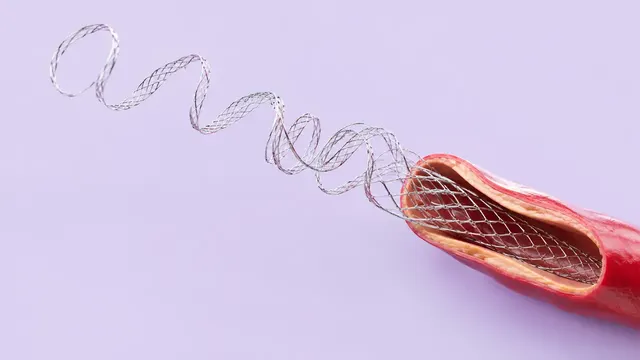
Coronary Angiography
Coronary angiography read: vessels, SYNTAX segments and stenosis with QCA evidence.
Cardiac13 sections
XA· cinefrom $0.12 / request
Peripheral and visceral DSA read: patency, stenoses, runoff, TASC II and GLASS stage, extravasation.
This model is not available to run yet. Vote to show interest and get an email when it opens.

Peripheral & Visceral Angiography reads digital subtraction angiography outside the heart and the head: aorto-iliac and lower-limb runoff studies, renal and mesenteric runs, and the runs recorded during embolisation for bleeding. Upload the whole study; its runs are grouped and read as one arterial map.
The result is a per-segment patency map from the iliac arteries to the pedal arch, with each stenosis and occlusion graded and measured, the number of patent tibial vessels, an anatomic stage for limb ischaemia, and, on visceral and trauma runs, the site and vessel of active contrast extravasation.
Decision support for physicians who plan or perform peripheral and visceral interventions. A structured patency map and stage help choose between endovascular treatment and bypass in chronic limb-threatening ischaemia, and a located extravasation documents the bleeding source on emergency and embolisation runs. Every lesion and bleeding site links to the run and frame that show it.
Interventional radiologists, vascular surgeons, interventional cardiologists who treat peripheral disease, and the residents and fellows who prepare the case. Vascular registries use the same structured staging.
| Requirement | Why |
|---|---|
| At least 16 frames | Opacification of each segment is followed over time |
| Not a rotational or cone-beam acquisition | These runs cannot be read as fixed projections and are excluded |
| Pixel spacing with source distances (or a magnification factor) | Lesion length and diameters in millimetres; without it, lengths stay in pixels and are flagged |
| Late frames down to the foot on runoff runs | Tibial runoff and the pedal arch fill late |
Stepping-table and stitched runs, patient motion and overlapping tibial vessels lower the confidence of the affected segments; those segments are named in the result instead of being graded silently.
The indication (claudication, chronic limb-threatening ischaemia, acute ischaemia, gastrointestinal or trauma
bleeding), the treated side and prior interventions can be passed in clinical_context.
One structured result per study with findings, measurements and stages, each linked to the run and frame it came from.
| Section | Content |
|---|---|
| Arterial segmentation | Arterial tree mask with segment labels: iliac, femoral, popliteal, tibial and pedal; renal and mesenteric on visceral runs |
| Stenoses and occlusions | Each lesion per arterial segment with percent stenosis or occlusion and its length |
| Runoff and anatomic staging | Patent tibial vessels to the foot, pedal arch, runoff score, TASC II class and GLASS stage |
| Active extravasation | Site and feeding vessel of contrast extravasation on visceral and trauma runs |
| Draft report | Opt-in (options.report): an English narrative that interprets the result for the reader (findings, impression, limitations); every number is checked against the findings |
The runoff score, TASC II class and GLASS stage are derived by a versioned rule from the measured lesions of the stenosis section, so each stage can be traced to the lesions that set it.
One run returns every section its input supports.
Coronary Angiography
Coronary angiography read: vessels, SYNTAX segments and stenosis with QCA evidence.
Cerebral DSA
Cerebral angiogram read: vessels, aneurysms, AV shunts, occlusions, stenosis and eTICI grading.
Intravascular OCT
IVOCT pullback read: lumen, plaque phenotype, calcium, stent sizing and strut-level stent analysis.
Intravascular Ultrasound (IVUS)
Vendor-neutral IVUS pullback read: lumen and EEM, plaque burden, calcium, sizing and stent result.
Cath-Lab Hemodynamics
Cath pressure tracings read: FFR and pullback pattern, valve gradients, right-heart values, QA.
LV Ventriculography & Aortography
LV gram and aortogram read: volumes, LVEF, regional wall motion and Sellers regurgitation grades.