Dermoscopy · Skin Lesion
Dermoscopic lesion read: ranked differential, malignancy risk, structures and 7-point score.
Skin6 sections
XC· photo
Rash and lesion photo read: top-5 differential, urgency, photo quality and severity scores.
This model is not available to run yet. Vote to show interest and get an email when it opens.
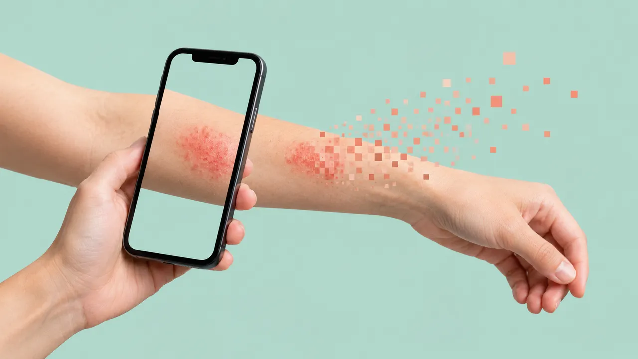
Clinical Skin Photo reads the everyday case: one to six phone or camera photos of a rash or a skin lesion, with a short history. The run first checks each photo for blur, lighting, framing, distance and obstruction, and says how to retake it when it cannot be graded. It then returns a top-5 differential over several hundred skin conditions, each coded in ICD-11 and SNOMED CT, with "cannot exclude" flags for conditions that should stay on the list.
An urgency class suggests the next step: urgent referral for suspected skin cancer, same-day review for a possible dermatological emergency, routine referral, or self-care. When the photos follow a scoring protocol (body regions, face views or scalp views), the run also returns the matching severity score: PASI for psoriasis, EASI for atopic dermatitis, an acne grade, VASI for vitiligo or SALT for alopecia areata.
Decision support for the clinician who sees the skin first, often without a dermatologist: a GP, an ED doctor, a nurse, or a teledermatology service sorting incoming cases. The severity scores support dermatologists and clinical trials that track treatment response with a fixed photo protocol.
GPs, emergency physicians, nurses, dermatologists, teledermatology providers and clinical-trial teams. Developers can call the run from a telehealth intake form to pre-fill the differential and urgency of each case for the dermatologist.
| Requirement | Why |
|---|---|
| A sharp, well-lit photo of the affected skin | Photos that cannot be graded are flagged with retake advice |
| An overview photo as well as a close-up | Distribution across the body is part of the differential |
| The full photo protocol for a severity score | Each score adds up area and intensity per region; missing regions leave it blank |
Age, sex, body site, duration, symptoms such as itch or pain, and how the rash has evolved, in clinical_context.
These fields change the differential materially, especially age, duration, itch and distribution.
One JSON result with a ranked, coded differential[], a management class and the severity sections that the photo
set supports.
| Section | Content |
|---|---|
| Photo quality | Gradable or not, with reasons (blur, lighting, framing, distance, obstruction) and retake advice |
| Differential diagnosis | Top-5 conditions with probabilities and "cannot exclude" flags, coded in ICD-10/ICD-11 and SNOMED CT |
| Urgency triage | Suspected skin cancer (urgent referral), possible dermatological emergency (same day), routine or self-care |
| PASI (psoriasis) | Erythema, induration, desquamation and area for each of four body regions, and the PASI total |
| EASI (atopic dermatitis) | Sign intensities and area per region with the EASI total; objective SCORAD optionally |
| Acne grade | Lesion counts by type and a severity grade |
| VASI (vitiligo) | Depigmented area per region and the VASI or facial VASI |
| SALT (alopecia areata) | Percentage of scalp hair loss and the SALT score |
| Draft report | Opt-in (options.report): an English narrative that interprets the result for the reader (findings, impression, limitations); every number is checked against the findings |
One run returns every section its input supports.
Dermoscopy · Skin Lesion
Dermoscopic lesion read: ranked differential, malignancy risk, structures and 7-point score.
Wound & Burn Photo
Wound and burn photo read: area in cm², tissue types, staging, foot flags and healing trend.
Total-Body Photography (mole mapping)
Total-body photo read: every lesion mapped, the most suspicious ranked, new and changed flagged.