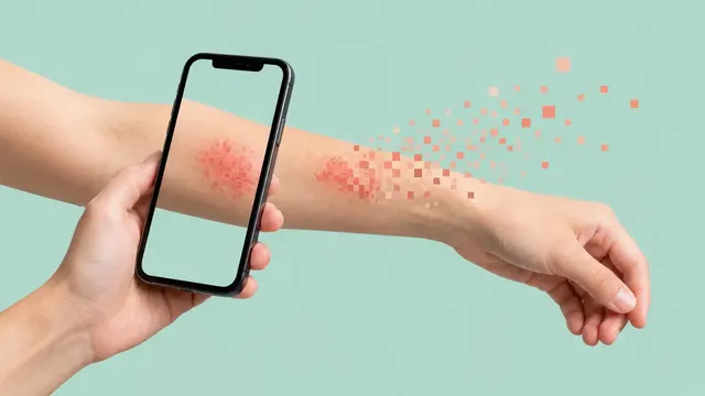
Clinical Skin Photo (rash or lesion)
Rash and lesion photo read: top-5 differential, urgency, photo quality and severity scores.
Skin9 sections
XC· photo
Dermoscopic lesion read: ranked differential, malignancy risk, structures and 7-point score.
This model is not available to run yet. Vote to show interest and get an email when it opens.

Dermoscopy reads one skin lesion seen through a dermatoscope. Upload one to four dermoscopic images of the lesion, polarised or non-polarised, with a clinical close-up and the patient's age, sex, body site and history if you have them. The run checks that each image is usable, then reads the lesion as a whole.
It returns a ranked differential (melanoma, naevus, basal cell carcinoma, squamous cell carcinoma or intraepidermal carcinoma, actinic keratosis, seborrhoeic keratosis or solar lentigo, dermatofibroma, vascular lesions and others) with calibrated probabilities and a malignancy probability. It names the dermoscopic structures it sees, in the terms of the International Dermoscopy Society (IDS), and derives a 7-point checklist score from them. A management class closes the read: excision or biopsy, referral, review by sequential dermoscopy at three months, or reassurance.
Decision support for the clinician holding the dermatoscope: a dermatologist, a dermatology resident, or a GP or nurse working in a skin-cancer or teledermoscopy pathway. The triage class is set to favour sensitivity, so a lesion is more likely to be sent for review than reassured. A reported history of change overrides a reassuring score.
Dermatologists, dermatology residents, GPs with a dermatoscope, skin-cancer clinic nurses and teledermatology services. Developers use the same run to add lesion analysis to a teledermoscopy platform or a clinic photo archive.
prior_files for the change section.| Requirement | Why |
|---|---|
| Lesion centred, at least about 600 px on the short side | Structures such as streaks, dots and vessels need resolution |
| Body site (supplied or estimated) | Acral, nail, mucosal and facial lesions follow their own pattern rules |
| Calibration sticker or ruler | Diameters and areas in mm; otherwise pixel values are returned and flagged |
| Little hair, ink, ruler marks or bubbles over the lesion | These artefacts are reported, and the quality check asks for a retake when they obscure the lesion |
Age, sex, body site, duration, symptoms (itch, bleeding), history of change, personal or family history of melanoma
and Fitzpatrick type in clinical_context. A clinical close-up and these fields refine the differential.
A JSON result with a lesion_id, a coded body site, the ranked differential[] and a management class.
| Section | Content |
|---|---|
| Differential diagnosis | Ranked diagnoses with calibrated probabilities over 8 diagnostic classes, coded in SNOMED CT and ICD-10/ICD-11 |
| Triage | Urgent referral or excision, routine referral, short-term sequential monitoring or reassurance, with the malignancy probability |
| Lesion segmentation | Lesion mask, area, maximum diameter, asymmetry and border-irregularity metrics |
| Dermoscopic structures | Pigment network, negative network, streaks, milia-like cysts, globules and dots, blue-white veil, regression structures and vessels, with masks and the 7-point checklist score |
| Change since prior | Registered comparison with the earlier image: growth, new structures, asymmetric enlargement and colour change, with an excise or keep-monitoring suggestion |
| Draft report | Opt-in (options.report): an English narrative that interprets the result for the reader (findings, impression, limitations); every number is checked against the findings |
One run returns every section its input supports.
Clinical Skin Photo (rash or lesion)
Rash and lesion photo read: top-5 differential, urgency, photo quality and severity scores.
Wound & Burn Photo
Wound and burn photo read: area in cm², tissue types, staging, foot flags and healing trend.
Total-Body Photography (mole mapping)
Total-body photo read: every lesion mapped, the most suspicious ranked, new and changed flagged.