Colonoscopy
Recorded colonoscopy read: polyps with optical diagnosis, BBPS, caecal intubation, withdrawal time.
GI7 sections
ES· video
Recorded laparoscopy review: phase timeline, critical view of safety, instruments and anatomy masks.
This model is not available to run yet. Vote to show interest and get an email when it opens.
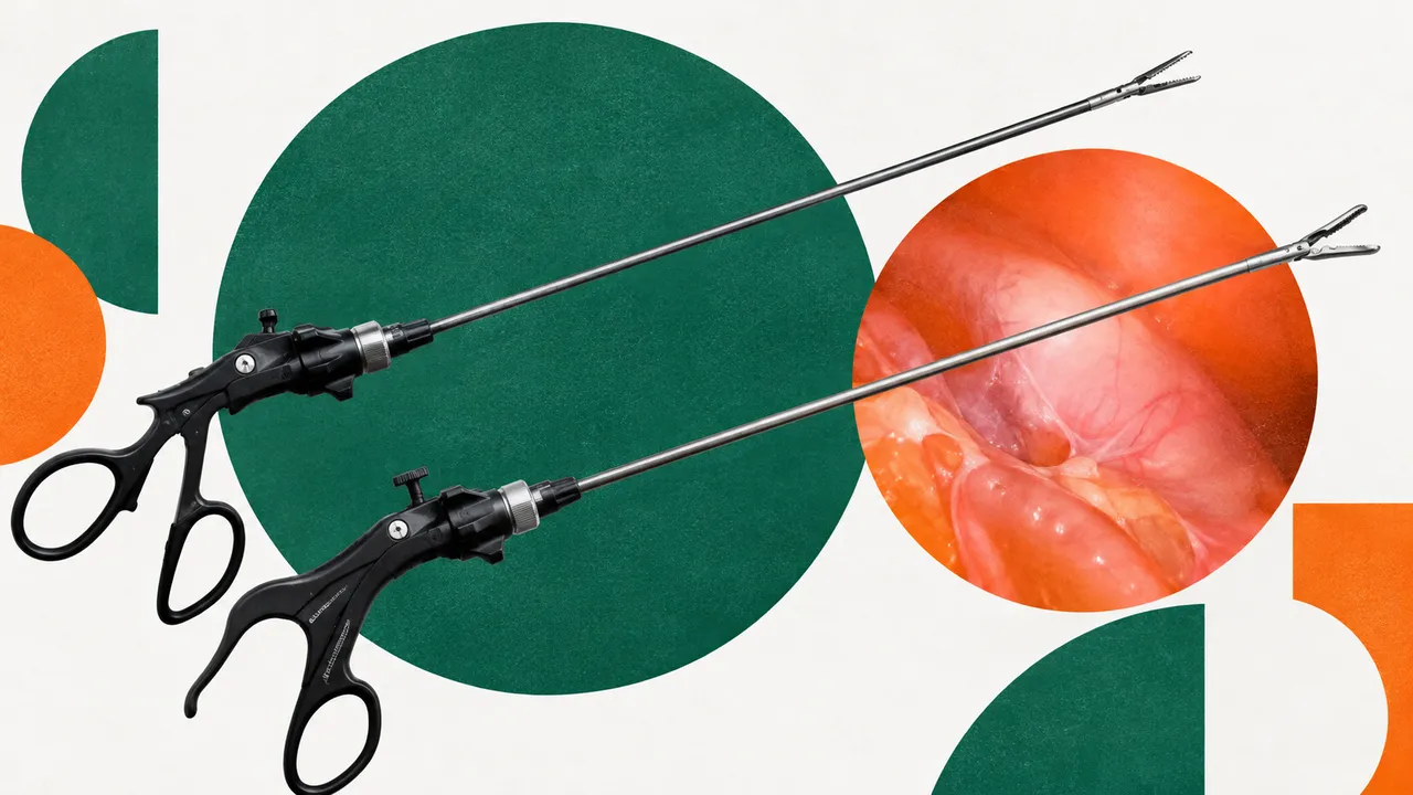
Laparoscopic Surgery Video reviews the recording of a laparoscopic operation after surgery. Cholecystectomy is one of the most common abdominal operations and the reference procedure for surgical-video analysis; the run reads it in full, and also gives the phase timeline of a laparoscopic hysterectomy.
For a cholecystectomy the result is a phase timeline with the duration of each phase, an assessment of the three Strasberg criteria of the critical view of safety with evidence frames, and the answer to the audit question: was the critical view achieved before the cystic structures were clipped? It also reports which instruments were in use and for how long, segments the hepatocystic-triangle anatomy, and lists key events such as bleeding, bile spillage and conversion.
Documentation, quality and teaching: a structured record of the operation, an audit of safe-cholecystectomy practice across a department, and structured feedback for residents on their own cases. Smoke, blood and unusual anatomy are the hard frames of the read.
General and hepatobiliary surgeons, surgical residents and their trainers, and quality and patient-safety teams. Developers and researchers use the run to index surgical video libraries by phase and instrument.
| Requirement | Why |
|---|---|
| The whole operation recorded | The phase timeline and durations need every phase |
| Dissection of the hepatocystic triangle in view before clipping | The CVS assessment is made on the frames before the first clip |
| Frame rate in the file | Phase durations and event times are reported in seconds |
The procedure and its setting (elective, or acute cholecystitis) can be passed in clinical_context.
| Section | Content |
|---|---|
| Surgical phases | Phase timeline with the start, end and duration of each phase |
| Critical view of safety | Each of the three Strasberg criteria per video segment, CVS achieved before clipping (yes or no) and evidence frames |
| Instruments | Tool presence per frame, tool segmentation and tracking, and usage time per instrument |
| Anatomy segmentation | Masks of gallbladder, cystic duct, cystic artery, liver and tools on key frames |
| Draft report | Opt-in (options.report): an English narrative that interprets the result for the reader (findings, impression, limitations); every number is checked against the findings |
Procedure.Fat and fibrous tissue are removed from the hepatocystic triangle; the lower third of the gallbladder is separated from the liver bed; and exactly two structures, the cystic duct and the cystic artery, are seen entering the gallbladder.
One run returns every section its input supports.
Colonoscopy
Recorded colonoscopy read: polyps with optical diagnosis, BBPS, caecal intubation, withdrawal time.
Capsule Endoscopy
Capsule study read: bleeding sources, lesions with Saurin relevance, transit times and key frames.
Upper GI Endoscopy (EGD)
Gastroscopy read: station completeness, LA grade, Barrett's extent and gastric neoplasia flags.
Bronchoscopy
Recorded bronchoscopy read: endobronchial tumours and mucosal abnormalities, located by airway.
Cystoscopy
Recorded cystoscopy read: papillary tumours and suspicious flat lesions, mapped with key frames.
Laryngoscopy and Stroboscopy
Laryngoscopy and stroboscopy read: vocal-fold lesions and glottal-area segmentation with waveform.