Colonoscopy
Recorded colonoscopy read: polyps with optical diagnosis, BBPS, caecal intubation, withdrawal time.
GI7 sections
ES· video
Gastroscopy read: station completeness, LA grade, Barrett's extent and gastric neoplasia flags.
This model is not available to run yet. Vote to show interest and get an email when it opens.
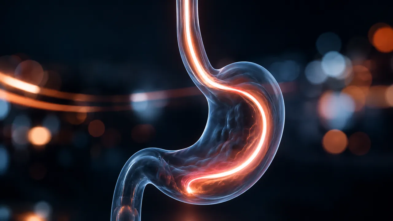
Upper GI Endoscopy reads a recorded oesophagogastroduodenoscopy, as video or as the still images saved during the examination. Missed early gastric cancer and Barrett's neoplasia are the main failures of EGD, and both are tied to an incomplete examination; the read checks the examination and the mucosa together.
The result lists which standard stations were seen and photographed, from the oesophagus and Z-line through the cardia, fundus, body, angulus, antrum and pylorus to the duodenum, and alerts on missing ones. In the distal oesophagus it detects erosive oesophagitis and assigns the Los Angeles grade. A Barrett's segment is measured as a Prague C&M estimate, with areas suspicious for dysplasia or early cancer marked for targeted biopsy. In the stomach, suspected early gastric cancer and adenomas are detected and delineated.
Decision support after the examination: a documented completeness check for quality audit, a second look at the oesophagogastric junction and stomach, and structured findings for the report. Lesion flags point to the frames that matter for biopsy planning.
Gastroenterologists, upper-GI surgeons, endoscopy trainees and units auditing photo-documentation. Developers use the run to add structured EGD findings and quality indicators to an endoscopy reporting system.
| Requirement | Why |
|---|---|
| Retroflexed view of the cardia and fundus | Completeness and the gastric read need the retroflexion station |
| Insufflated, washed oesophagus | Mucus, bubbles and collapsed lumen hide erosions and Barrett's tongues |
| Clear view of the Z-line and the top of the gastric folds | Separates short-segment Barrett's from an irregular Z-line and anchors Prague C&M |
| Close views of suspicious areas | Neoplasia flags need near views, ideally with image enhancement |
Indication (dyspepsia, reflux, Barrett's surveillance, bleeding), known Barrett's length and prior histology can be
passed in clinical_context.
| Section | Content |
|---|---|
| Examination completeness | Stations seen and photographed, with a missing-station alert |
| Oesophagitis (LA grade) | Erosive oesophagitis present or absent and Los Angeles grade A–D |
| Barrett's oesophagus | Prague C&M estimate and neoplasia flags for targeted biopsy |
| Gastric neoplasia | Detected and delineated suspected early gastric cancer or adenoma, with Paris type |
| Draft report | Opt-in (options.report): an English narrative that interprets the result for the reader (findings, impression, limitations); every number is checked against the findings |
Procedure and DiagnosticReport.Prague C is the circumferential extent and M the maximum extent of columnar mucosa above the gastro-oesophageal junction, in centimetres. From video, both are estimated without a calibrated scale.
One run returns every section its input supports.
Colonoscopy
Recorded colonoscopy read: polyps with optical diagnosis, BBPS, caecal intubation, withdrawal time.
Capsule Endoscopy
Capsule study read: bleeding sources, lesions with Saurin relevance, transit times and key frames.
Bronchoscopy
Recorded bronchoscopy read: endobronchial tumours and mucosal abnormalities, located by airway.
Cystoscopy
Recorded cystoscopy read: papillary tumours and suspicious flat lesions, mapped with key frames.
Laparoscopic Surgery Video
Recorded laparoscopy review: phase timeline, critical view of safety, instruments and anatomy masks.
Laryngoscopy and Stroboscopy
Laryngoscopy and stroboscopy read: vocal-fold lesions and glottal-area segmentation with waveform.