
Breast Tomosynthesis
DBT read: lesions located in 3D with their best slice, exam score, recall and BI-RADS.
Breast2 sections
MG· image-2d-multiview
Mammogram read: suspicion score, recall, BI-RADS, density, located lesions and risk.
This model is not available to run yet. Vote to show interest and get an email when it opens.
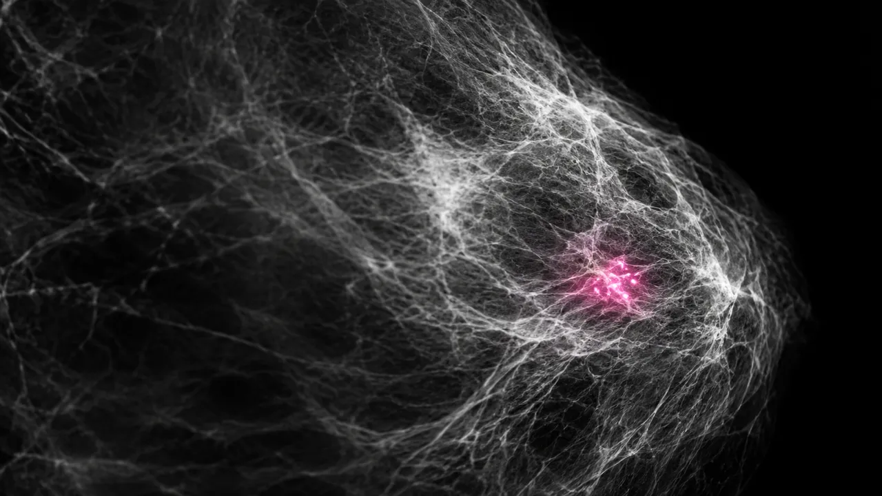
Mammogram (2D) reads a full-field digital or synthesized 2D mammogram as a second reader. Upload the four standard views
(left and right CC and MLO), any additional diagnostic views and the prior examinations. Set the exam_context option
to screening or diagnostic: a screening read returns recall or no recall with BI-RADS 0, 1 or 2, and a diagnostic read
returns a BI-RADS 1–6 assessment for each lesion.
Every read carries an exam suspicion score from 0 to 100, a BI-RADS assessment per breast and for the exam, and the density category. Masses, calcification clusters, architectural distortion and asymmetries are boxed on each view, matched between CC and MLO, and located by laterality, clock face, depth and distance from the nipple, each with BI-RADS lexicon descriptors and its own score. Positioning quality, change against priors, image-based risk and arterial calcification complete the result.
Decision support for the radiologist reading screening or diagnostic mammograms, including general radiologists without breast fellowship training in high-volume programmes: a second read that marks and scores every lesion, a density category for notification and supplemental-screening decisions, and positioning feedback for technologists.
Breast and general radiologists, screening programmes, mammography technologists, breast surgeons and high-risk clinics. Developers can add a second-reader score, density and positioning checks to a PACS, screening worklist or reporting system.
prior_files; strongly recommended.| Requirement | Why |
|---|---|
| All four standard views | Lesions are matched across CC and MLO, and the risk section needs the complete set |
| For Presentation images for the risk section | The risk estimate is computed on For Presentation FFDM |
ImageLaterality, ViewPosition and view code sequences |
Views and laterality are verified before reading |
BreastImplantPresent |
Implant and implant-displaced views are read differently |
| Pixel spacing | Lesion size and distance from the nipple in mm |
Incomplete studies, implants and differences between synthesized 2D and FFDM appearance are flagged in the result.
Age, risk factors and the clinical indication (lump, nipple discharge, follow-up) in clinical_context; the
exam_context option selects screening or diagnostic logic.
A JSON result with the exam score, recall decision, BI-RADS assessments and the sections below.
| Section | Content |
|---|---|
| Positioning quality | PGMI grade per view, posterior nipple line CC versus MLO, pectoral muscle, nipple in profile, inframammary fold, skin folds, motion and retake advice |
| Breast density | BI-RADS density a–d per breast and exam, dense-tissue percentage and a dense or not-dense flag |
| Lesions | Masses, calcification clusters, architectural distortion and asymmetries per view, with malignancy score, CC–MLO match and clock-face location |
| Calcifications | Microcalcification clusters with masks, BI-RADS morphology and distribution, and a suspicion score |
| Comparison with priors | New or enlarging masses, new calcifications, developing asymmetry, density change and stability over two years or more |
| Breast cancer risk | 1- to 5-year risk, a risk band and a supplemental-screening suggestion |
| Breast arterial calcification | Presence and severity grade, with a cardiovascular-risk flag |
| Draft report | Opt-in (options.report): an English narrative that interprets the result for the reader (findings, impression, limitations); every number is checked against the findings |
One run returns every section its input supports.
Breast Tomosynthesis
DBT read: lesions located in 3D with their best slice, exam score, recall and BI-RADS.
Contrast-Enhanced Mammography
CEM read: enhancing masses and non-mass enhancement, BPE level and BI-RADS by the CEM lexicon.