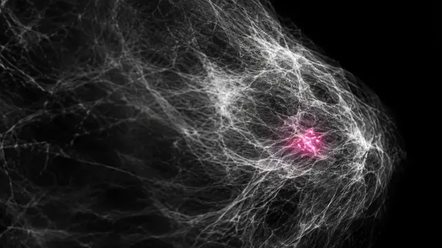
Mammogram (2D)
Mammogram read: suspicion score, recall, BI-RADS, density, located lesions and risk.
Breast8 sections
MG· image-2d-multiview
CEM read: enhancing masses and non-mass enhancement, BPE level and BI-RADS by the CEM lexicon.
This model is not available to run yet. Vote to show interest and get an email when it opens.

Contrast-Enhanced Mammography reads a CEM study (also called contrast-enhanced spectral mammography, CESM). After an iodinated contrast injection, each view is acquired as a low-energy image, which looks like a standard mammogram, and a recombined image that shows iodine uptake. The run reads both images of every view together.
On the recombined images it finds enhancing masses and non-mass enhancement, outlines them, grades how strongly they enhance and scores each for malignancy, using the matching low-energy image to place them in the breast. It grades background parenchymal enhancement (BPE) for each breast with its symmetry, because marked BPE can hide a lesion. A BI-RADS assessment closes the read, using the CEM lexicon of BI-RADS v2025.
Decision support for radiologists using CEM to work up a recalled or symptomatic patient, to stage a known cancer, or as an alternative where breast MRI is not available. Each enhancing lesion comes with its mask, enhancement level and malignancy score, and the BI-RADS assessment carries the management that follows.
Breast radiologists, and general radiologists in centres that are adopting CEM without MRI access; breast surgeons and oncologists planning surgery. Developers can add structured CEM reads to a breast imaging PACS or reporting system.
ImageType and series descriptions, so the pairs do not need to be labelled.| Requirement | Why |
|---|---|
| Paired low-energy and recombined images for each view | Enhancement is read on the recombined image and located on the low-energy image |
| CC and MLO of both breasts | Lesions are matched across views, and BPE is graded and compared per breast |
ImageLaterality and view codes |
Each pair is assigned to the right breast and projection |
| Pixel spacing | Lesion size in mm |
Vendor differences in recombination and recombination artefacts (rim and ripple) are recognised and flagged where they affect the read.
Indication (recall, symptoms, staging of a known cancer), age and prior imaging in
clinical_context and prior_files.
A JSON result with a BI-RADS assessment and the two CEM sections, with masks and boxes on the recombined images.
| Section | Content |
|---|---|
| Enhancing lesions | Enhancing masses and non-mass enhancement with boxes, masks, enhancement level and malignancy score |
| Background parenchymal enhancement | BPE level (minimal, mild, moderate or marked) per breast and its symmetry |
| Draft report | Opt-in (options.report): an English narrative that interprets the result for the reader (findings, impression, limitations); every number is checked against the findings |
One run returns every section its input supports.