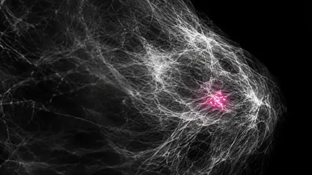
Mammogram (2D)
Mammogram read: suspicion score, recall, BI-RADS, density, located lesions and risk.
Breast8 sections
MG· image-2d-multiview
DBT read: lesions located in 3D with their best slice, exam score, recall and BI-RADS.
This model is not available to run yet. Vote to show interest and get an email when it opens.

Breast Tomosynthesis reads a digital breast tomosynthesis (DBT) study: one reconstructed stack of thin slices for each view, usually with synthesized 2D images and sometimes with conventional FFDM views. DBT finds more cancers and causes fewer recalls than 2D mammography, but a radiologist has to scroll through 50 to 90 slices per view, which roughly doubles the reading time.
The run searches every slice of every stack. Each mass, area of architectural distortion and calcification cluster is returned as a 3D box with its slice range, the single best slice to look at, and its own score. The exam-level read adds a suspicion score, a recall recommendation and a BI-RADS assessment, so the radiologist can open the study at the slices that matter.
Decision support for the radiologist reading DBT in screening or diagnostic work: a second read that points to the slices and regions that matter, and a structured exam assessment with a recall recommendation. Every lesion comes with its location in the stack.
Breast and general radiologists reading tomosynthesis, screening programmes that have moved from 2D to DBT, and breast clinics. Developers can call the run from a PACS or screening worklist to pre-mark key slices before the reader opens the study.
prior_files, as DBT or 2D examinations.A DBT study is large, typically 1 to 3 GB, with 50 to 90 slices per view at about 1 mm spacing.
| Requirement | Why |
|---|---|
| A tomosynthesis stack for each standard view | Lesions are detected in 3D and matched between CC and MLO |
ImageLaterality and view codes |
Each stack is assigned to the correct breast and projection |
| Slice spacing and pixel spacing | Slice ranges and lesion sizes in mm |
BreastImplantPresent |
Implant and implant-displaced views are recognised |
Image intensities are normalised per vendor before reading; incomplete studies are flagged.
Screening or diagnostic exam_context, age, risk factors and the clinical indication in clinical_context.
A JSON result with the exam suspicion score, recall decision, BI-RADS assessment and a lesion list carrying slice coordinates.
| Section | Content |
|---|---|
| Lesions in 3D | Masses, architectural distortion and calcification clusters as 3D boxes with slice range, best slice, laterality, clock-face location and per-lesion score |
| Draft report | Opt-in (options.report): an English narrative that interprets the result for the reader (findings, impression, limitations); every number is checked against the findings |
Breast Tomosynthesis shares its lesion lexicon, location fields and assessment logic with Mammogram (2D), so DBT and 2D reads of the same patient line up when the reader compares them over time.
One run returns every section its input supports.
Mammogram (2D)
Mammogram read: suspicion score, recall, BI-RADS, density, located lesions and risk.
Contrast-Enhanced Mammography
CEM read: enhancing masses and non-mass enhancement, BPE level and BI-RADS by the CEM lexicon.