Chest CT
Chest CT read: nodules with Fleischner or Lung-RADS, PE with RV strain, lungs, pleura and ribs.
Chest16 sections
CT· volume
Cardiac CT read: Agatston calcium, CAD-RADS 2.0 stenosis, plaque, chambers and TAVI measurements.
This model is not available to run yet. Vote to show interest and get an email when it opens.
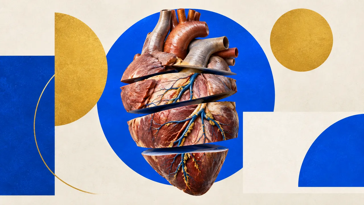
Cardiac CT reads ECG-gated heart studies: the non-contrast calcium-score scan, coronary CT angiography for chest pain, and the structural protocols for transcatheter aortic valve implantation (TAVI) and left atrial appendage occlusion (LAAO). The run tells these protocols and cardiac phases apart and computes what each one supports.
From a calcium scan it returns the Agatston score per coronary artery with its CAC-DRS category and the epicardial fat volume. From a CCTA it returns the labelled coronary tree, stenosis per segment with a CAD-RADS 2.0 category and modifiers, plaque volumes by composition, chamber volumes and CT-FFR per lesion. A TAVI or LAAO study adds the device-sizing measurements the heart team needs.
Decision support for the reader of a cardiac CT and for the team that acts on it: a CAD-RADS category with the segment measurements behind it, and TAVI or LAAO sizing values taken at defined landmarks. When a patient goes on to invasive angiography, the Coronary Angiography model reads the cath-lab study.
Cardiac radiologists, imaging and interventional cardiologists, structural-heart teams and cardiac surgeons. Developers add calcium scoring, CAD-RADS or TAVI sizing to a reporting system or a structural-heart planning workflow.
| Requirement | Why |
|---|---|
| ECG gating and cardiac phase fields | Phase detection and selection of the motion-free reconstruction |
| Rescale Slope and Intercept | Calcium scoring and plaque composition are both defined in Hounsfield units |
| Full coverage of the coronary tree | Segments outside the volume are reported as not evaluable |
| Several phases of the cardiac cycle | Ejection fraction is computed only from multiphase data |
| For TAVI, run-off from the aortic root to the femoral arteries | Iliofemoral minimal lumen diameters for the access route |
Motion and step artefacts, calcium blooming and coronary stents are flagged per segment.
The indication (chest pain, pre-TAVI, pre-LAAO, risk assessment), known stents or bypass grafts and the intended device
go in clinical_context. The targets option extends the calcium score to the aortic valve and mitral annulus.
A sectioned JSON result built on the coronary segment model; every stenosis and plaque value points to its segment.
| Section | Content |
|---|---|
| Calcium score | Agatston, volume and mass per vessel (LM, LAD, LCX, RCA) and in total, with the CAC-DRS category |
| Coronary tree segmentation | Coronary mask, centrelines and segment labels |
| CAD-RADS stenosis | Stenosis grade per segment, plaque burden and modifiers (N, HRP, S, G, E), category and management |
| Coronary plaque | Calcified, non-calcified and low-attenuation plaque volumes, total plaque volume, high-risk features |
| CT-FFR | Lesion-specific fractional flow reserve computed from the CCTA |
| Cardiac chambers | LA, LV, RA, RV, LV myocardium, aorta and pulmonary artery; volumes, LV mass and EF when multiphase |
| TAVI planning | Annulus area, perimeter and diameters, LVOT, sinus and STJ diameters, coronary ostial heights, cusp calcium, iliofemoral minimal lumen diameters |
| LAA occlusion planning | Appendage morphology, ostium and landing-zone diameters, depth, filling-defect flag (thrombus or slow flow) |
| Epicardial fat | Epicardial adipose tissue volume and mean attenuation inside the pericardium |
| Draft report | Opt-in (options.report): an English narrative that interprets the result for the reader (findings, impression, limitations); every number is checked against the findings |
One run returns every section its input supports.
Chest CT
Chest CT read: nodules with Fleischner or Lung-RADS, PE with RV strain, lungs, pleura and ribs.
Head CT
Head CT read: bleed volumes, ASPECTS, vessel occlusion, perfusion core and skull fractures.
Abdomen & Pelvis CT
Abdomen-pelvis CT read: acute abdomen flags, organ lesions with management, stones and trauma.
Spine CT
Spine CT read: every vertebra labelled, fractures with AO Spine type, alignment and canal size.
CT Angiography (Aorta, Carotid, Run-off)
Arterial CTA read: aortic diameters, dissection and aneurysm, endoleak, carotid and leg stenoses.
Head & Neck CT
Neck, sinus and temporal-bone CT read: nodes by level, Lund-Mackay, airway, abscess and tumour.