Chest CT
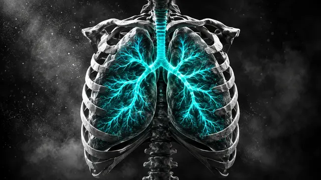
Chest CT read: nodules with Fleischner or Lung-RADS, PE with RV strain, lungs, pleura and ribs.
Chest16 sections
CT· volume
Neck, sinus and temporal-bone CT read: nodes by level, Lund-Mackay, airway, abscess and tumour.
This model is not available to run yet. Vote to show interest and get an email when it opens.
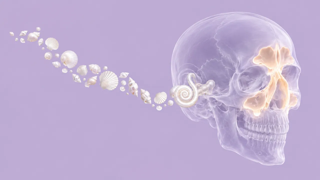
Head & Neck CT covers the ENT family of CT protocols: the contrast-enhanced neck CT, the paranasal sinus CT and the high-resolution temporal-bone CT. Upload any of them; the run identifies the protocol and reads the sections that protocol supports, so a sinus CT returns a Lund-Mackay score and a neck CT returns nodes, abscesses and tumour findings.
On a contrast neck CT the run maps lymph nodes to levels I–VII with short axis, necrosis and extranodal-extension signs, detects deep neck abscesses and a primary tumour, and measures the upper airway. On a sinus CT it grades opacification of every sinus and the ostiomeatal complex. On a temporal-bone CT it reads the ossicles, otic capsule, scutum and semicircular canals for erosion, cholesteatoma signs and dehiscence. Facial fractures are read by the Head CT model, not here.
Decision support for the clinician who reads or acts on a head and neck CT: the ENT surgeon planning sinus surgery, the emergency physician with a febrile patient and neck swelling at night, the oncologist following treated cancer. Every finding carries its measurements.
Head and neck radiologists, ENT and maxillofacial surgeons, radiation and medical oncologists, and emergency physicians. Developers use the run to bring structured nodal levels and sinus scores into an ENT clinic system or a tumour-board tool.
prior_files when post-treatment change is in question.| Requirement | Why |
|---|---|
| Intravenous contrast for neck sections | Node necrosis, abscess rim and tumour enhancement need it |
| Neck or chest coverage for the airway | Lumen area and stenosis length are measured along the covered airway |
| Sinus CT covering all paranasal sinuses | Lund-Mackay sums every sinus on both sides |
| Thin bone-kernel reconstruction for temporal bone | Ossicles and canal walls are sub-millimetre structures |
Dental amalgam streaks and swallowing motion are reported as quality flags.
The indication (infection, staging, post-treatment surveillance, chronic rhinosinusitis, hearing loss) and the
treatment history go in clinical_context. For a treated neck, findings are framed in NI-RADS post-treatment
terms.
A sectioned JSON result with findings, measurements and classifications, plus DICOM objects for the masks.
| Section | Content |
|---|---|
| Cervical lymph nodes | Nodes per level I–VII: short axis, necrosis, extranodal-extension signs and a Node-RADS score |
| Primary tumour | Oral cavity, oropharynx, larynx or nasopharynx tumour: segmentation, size and invasion flags |
| Deep neck abscess | Peritonsillar, parapharyngeal or retropharyngeal abscess: size, airway compromise, mediastinal extension |
| Upper airway | Lumen segmentation, minimal cross-sectional area, stenosis length and extrinsic compression |
| Lund-Mackay sinus score | Opacification 0, 1 or 2 per sinus, ostiomeatal-complex patency, total 0–24 |
| Temporal bone | Ossicular chain and otic capsule: scutum erosion, cholesteatoma signs, canal dehiscence |
| Draft report | Opt-in (options.report): an English narrative that interprets the result for the reader (findings, impression, limitations); every number is checked against the findings |
On each side, the maxillary, anterior ethmoid, posterior ethmoid, sphenoid and frontal sinuses are graded 0 (clear), 1 (partial) or 2 (total opacification), and the ostiomeatal complex 0 (patent) or 2 (obstructed). Both sides together give a Lund-Mackay total from 0 to 24. Every node is assigned to one of levels I–VII and measured in short axis, then scored with Node-RADS 1.0.
One run returns every section its input supports.
Chest CT
Chest CT read: nodules with Fleischner or Lung-RADS, PE with RV strain, lungs, pleura and ribs.
Head CT
Head CT read: bleed volumes, ASPECTS, vessel occlusion, perfusion core and skull fractures.
Abdomen & Pelvis CT
Abdomen-pelvis CT read: acute abdomen flags, organ lesions with management, stones and trauma.
Cardiac CT
Cardiac CT read: Agatston calcium, CAD-RADS 2.0 stenosis, plaque, chambers and TAVI measurements.
Spine CT
Spine CT read: every vertebra labelled, fractures with AO Spine type, alignment and canal size.
CT Angiography (Aorta, Carotid, Run-off)
Arterial CTA read: aortic diameters, dissection and aneurysm, endoleak, carotid and leg stenoses.