Chest CT
Chest CT read: nodules with Fleischner or Lung-RADS, PE with RV strain, lungs, pleura and ribs.
Chest16 sections
CT· volume
Spine CT read: every vertebra labelled, fractures with AO Spine type, alignment and canal size.
This model is not available to run yet. Vote to show interest and get an email when it opens.
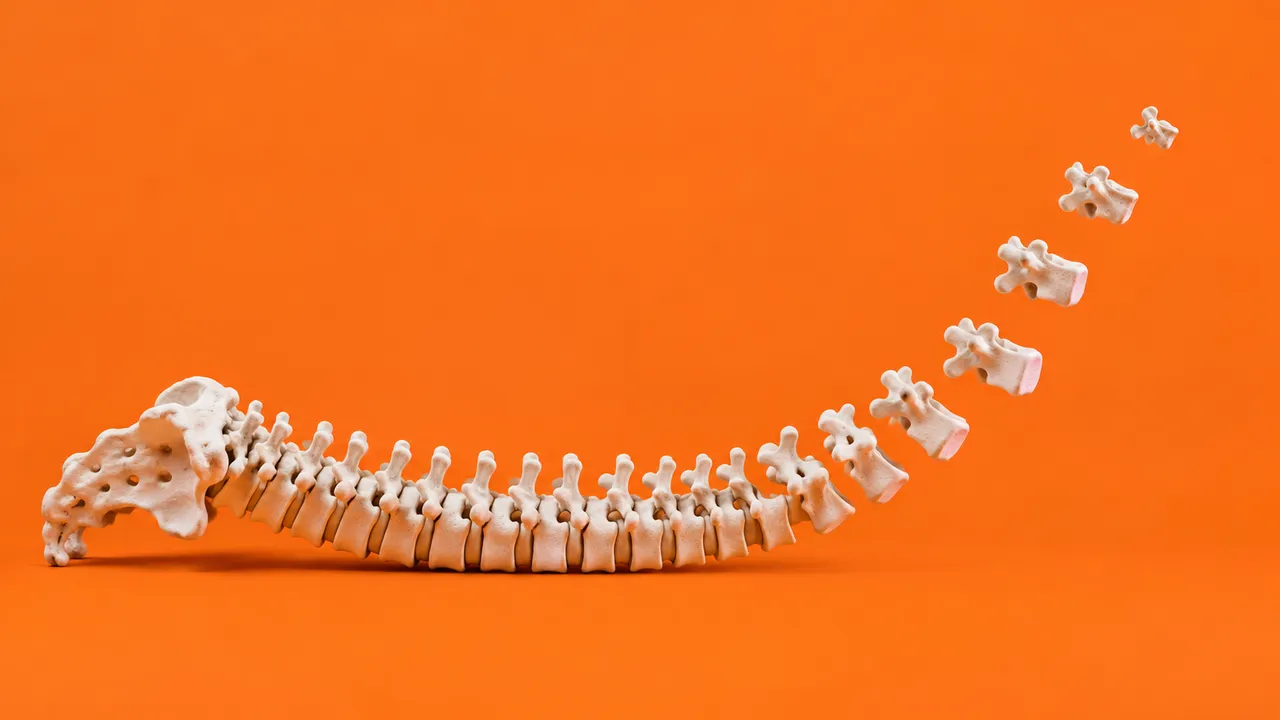
Spine CT reads the cervical, thoracic and lumbar spine, either from a dedicated spine CT or from the spine reformats of a trauma or body CT. The run first segments and labels every vertebra from C1 to L5 and the sacrum, with the vertebral bodies and intervertebral discs, so that each later finding is tied to a named level.
On that labelled spine it looks for fractures and describes their morphology (compression, burst, distraction, translation) with an AO Spine thoracolumbar type at thoracic and lumbar levels, assesses alignment and canal compromise, and measures the canal and foramina at every level for degenerative narrowing. In the emergency department this is the read where a missed cervical fracture costs the most; in clinic it is a measured map of stenosis before surgery.
Decision support for the clinician clearing the spine after trauma, often the emergency physician or radiology resident reading a head and cervical spine CT together at night, and for the spine surgeon planning decompression or fixation. Each fracture and narrowing is reported at a named level, with its measurements.
Emergency and musculoskeletal radiologists, emergency physicians, trauma teams, spine surgeons, neurosurgeons and orthopaedic surgeons. Developers use the labelled vertebrae and level-based findings in trauma worklists, surgical planning software or spine registries.
| Requirement | Why |
|---|---|
| Bone-kernel reconstruction | Fracture lines and cortical breaks are read on the sharp kernel |
| Thin axial slices allowing sagittal reformats | Height loss, alignment and canal measurements are sagittal |
| Image Position and Orientation (Patient) | Slices are ordered geometrically, which matters for reformats of long body scans |
Transitional anatomy (lumbarised or sacralised segments, cervical ribs) is flagged when detected, because it can shift level numbering. Shoulder artefact at C7–T1, osteophytes, vascular channels and Mach bands are handled as quality flags and known mimics.
The mechanism of injury, neurological symptoms, known previous fractures or fixation hardware and the clinical question
(trauma clearance or degenerative work-up) go in clinical_context.
A sectioned JSON result in which every finding carries its vertebral level, with masks in DICOM SEG.
| Section | Content |
|---|---|
| Spine fractures | Fracture per vertebral level, morphology (compression, burst, distraction, translation), AO Spine type at thoracolumbar levels, canal compromise |
| Vertebrae and discs | Masks and labels for C1–L5 and the sacrum, vertebral bodies and intervertebral discs |
| Canal and foraminal stenosis | Canal anteroposterior diameter and area per level, foraminal narrowing, disc-height loss, facet hypertrophy flags |
| Draft report | Opt-in (options.report): an English narrative that interprets the result for the reader (findings, impression, limitations); every number is checked against the findings |
The trauma read also states alignment and canal compromise at each injured level.
Old healed fractures, osteophytes and degenerative endplate change can resemble acute injury, and vascular channels or Mach bands can mimic a fracture line. Each fracture therefore comes with its level, key image and morphology. For a head and cervical spine trauma study, the Head CT model reads the brain and skull in a separate run.
One run returns every section its input supports.
Chest CT
Chest CT read: nodules with Fleischner or Lung-RADS, PE with RV strain, lungs, pleura and ribs.
Head CT
Head CT read: bleed volumes, ASPECTS, vessel occlusion, perfusion core and skull fractures.
Abdomen & Pelvis CT
Abdomen-pelvis CT read: acute abdomen flags, organ lesions with management, stones and trauma.
Cardiac CT
Cardiac CT read: Agatston calcium, CAD-RADS 2.0 stenosis, plaque, chambers and TAVI measurements.
CT Angiography (Aorta, Carotid, Run-off)
Arterial CTA read: aortic diameters, dissection and aneurysm, endoleak, carotid and leg stenoses.
Head & Neck CT
Neck, sinus and temporal-bone CT read: nodes by level, Lund-Mackay, airway, abscess and tumour.