Head CT
Head CT read: bleed volumes, ASPECTS, vessel occlusion, perfusion core and skull fractures.
Neuro12 sections
CT· volume
Chest CT read: nodules with Fleischner or Lung-RADS, PE with RV strain, lungs, pleura and ribs.
This model is not available to run yet. Vote to show interest and get an email when it opens.
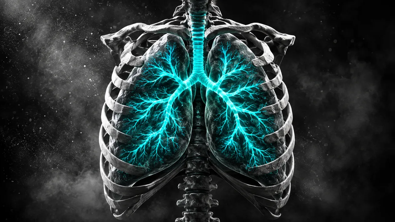
Chest CT reads every chest protocol with one model: non-contrast and contrast-enhanced CT, CT pulmonary angiography, low-dose lung-cancer screening and high-resolution CT for interstitial lung disease. The run works out the protocol from the kernel, slice thickness, contrast timing and pulmonary-artery enhancement, then runs the sections that protocol supports.
The result covers the whole chest. Nodules come with size, volume, type and management per the 2017 Fleischner guidelines, or a Lung-RADS v2022 category on a screening scan. On a CTPA, emboli are reported with the most proximal clot level, clot burden and right-ventricular strain. Parenchyma (emphysema, fibrosis, infection), airways, pleura, ribs and coronary calcium complete the read, with lungs, lobes and pulmonary vessels segmented underneath.
Decision support for chest CT readers: the on-call resident with a night-time CTPA, the screening-programme radiologist assigning Lung-RADS, the pulmonologist following fibrosis or emphysema. Every finding carries its mask and measurements.
Thoracic and emergency radiologists, pulmonologists, emergency physicians, thoracic surgeons and lung-screening programmes. Developers use the run to add nodule management, PE triage or quantitative lung metrics to a reading platform or registry.
prior_files: nodule growth and the Lung-RADS category of a follow-up screen use it.| Requirement | Why |
|---|---|
| Slices of 2.5 mm or thinner, lung kernel preferred | Nodule detection, volume and type |
| Pulmonary-artery enhancement on CTPA | A contrast-adequacy check runs before any embolus is called |
| Prior low-dose scan for a follow-up screen | Lung-RADS growth criteria compare against it; a baseline screen needs none |
| Inspiratory non-contrast series | LAA-950 is defined on inspiratory, unenhanced lung density |
| Prone and expiratory HRCT series, when acquired | Prone images separate dependent atelectasis from fibrosis; expiratory images show air trapping |
| Bone kernel | Rib fracture detection and numbering |
Respiratory motion, mixing artefact and screening-dose noise are reported as quality flags.
The screening or diagnostic setting and the clinical question (suspected PE, staging, dyspnoea) go in
clinical_context. With the detected protocol, they decide between Fleischner and Lung-RADS management.
One sectioned JSON result. Each nodule and clot has its own mask, measurements and lobe location.
| Section | Content |
|---|---|
| Emergency triage | Flags for PE, pneumothorax, aortic dissection or large aneurysm, large pleural or pericardial effusion |
| Pulmonary embolism | Clot detection, most proximal level (saddle to subsegmental), clot masks and burden, RV-strain flags, contrast adequacy |
| RV/LV ratio | Maximal RV and LV diameters, the ratio and the main pulmonary-artery diameter |
| Lung nodules | Detection, 3D segmentation, diameter and volume, solid, part-solid or ground-glass, malignancy score, follow-up |
| Lung-RADS | Growth against the prior, category, management and the S modifier for significant incidental findings |
| Lung-cancer risk | Predicted risk of lung cancer over 1 to 6 years from a screening scan |
| Emphysema | Lung and lobe segmentation, LAA-950 percentage per lobe and distribution |
| Interstitial lung disease | Reticulation, honeycombing, traction bronchiectasis and ground glass, extent and UIP-pattern category |
| Infection extent | Consolidation and ground-glass segmentation, percentage involvement per lobe and severity |
| Airways | Airway tree to sub-segmental level, wall thickness, broncho-arterial ratio, bronchiectasis and mucus plugging |
| Pleural and pericardial effusion | Segmentation, volume in millilitres and density, with a haemothorax suspicion flag |
| Pneumothorax | Side, volume and tension signs such as mediastinal shift |
| Rib fractures | Detected fractures with rib number, type (displaced, non-displaced, buckle, segmental) and flail-segment flag |
| Coronary calcium | Agatston-equivalent score and ordinal category on non-gated CT, including low-dose scans |
| Lung, lobe and vessel segmentation | Lungs, five lobes, pulmonary arteries and veins and airways, with volumes |
| Draft report | Opt-in (options.report): an English narrative that interprets the result for the reader (findings, impression, limitations); every number is checked against the findings |
One run returns every section its input supports.
Head CT
Head CT read: bleed volumes, ASPECTS, vessel occlusion, perfusion core and skull fractures.
Abdomen & Pelvis CT
Abdomen-pelvis CT read: acute abdomen flags, organ lesions with management, stones and trauma.
Cardiac CT
Cardiac CT read: Agatston calcium, CAD-RADS 2.0 stenosis, plaque, chambers and TAVI measurements.
Spine CT
Spine CT read: every vertebra labelled, fractures with AO Spine type, alignment and canal size.
CT Angiography (Aorta, Carotid, Run-off)
Arterial CTA read: aortic diameters, dissection and aneurysm, endoleak, carotid and leg stenoses.
Head & Neck CT
Neck, sinus and temporal-bone CT read: nodes by level, Lund-Mackay, airway, abscess and tumour.