Chest CT
Chest CT read: nodules with Fleischner or Lung-RADS, PE with RV strain, lungs, pleura and ribs.
Chest16 sections
CT· volume
Pelvis and extremity CT read: fractures with fragments, 3D bone models and Bone-RADS lesions.
This model is not available to run yet. Vote to show interest and get an email when it opens.
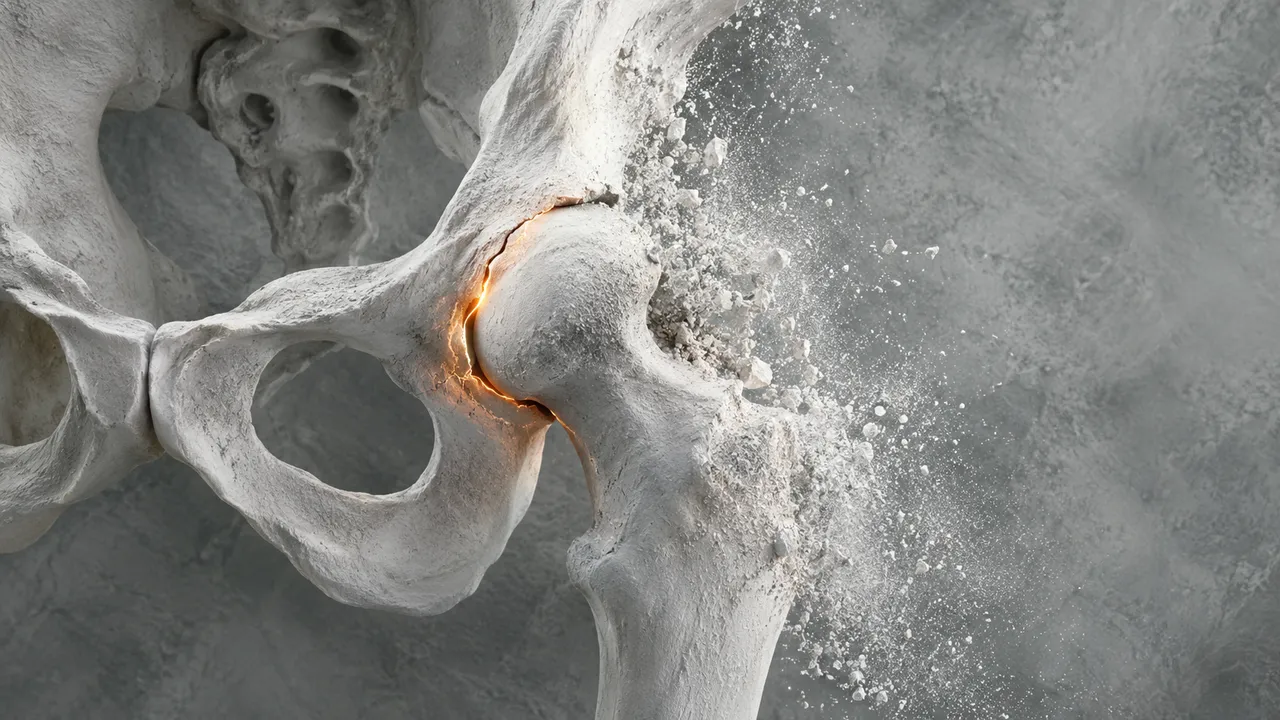
Musculoskeletal CT reads CT of the pelvis, acetabulum and limbs, the scans ordered for complex fractures, for planning their fixation and for characterising a bone lesion. The run segments every bone in the field of view, together with any implant, and exports each one as a surface mesh that planning software and 3D printers can open.
On those masks it detects fractures, splits each fractured bone into labelled fragments and measures their displacement, which turns a stack of axial slices into a fragment map a surgeon can rotate. Focal bone lesions are found, described as lytic, sclerotic or mixed with their aggressiveness features, and given the management category of Bone-RADS.
Decision support and planning input for orthopaedic and trauma surgeons and for the emergency physicians who see these injuries first: where the fracture runs, how many fragments there are and how far they have moved. For incidental bone lesions it supplies a structured, guideline-based next step.
Orthopaedic trauma surgeons, pelvic and acetabular surgeons, hand and foot surgeons, musculoskeletal radiologists, emergency physicians and 3D-printing labs in hospitals. Developers use the meshes and fragment labels in surgical planning, patient-specific guide or implant design software.
| Requirement | Why |
|---|---|
| Thin slices | Surface meshes and fragment boundaries follow the voxel size; thick slices are flagged for partial volume |
| Rescale Slope and Intercept | Bone, metal and lesion matrix are separated in Hounsfield units |
| The whole bone of interest in the field of view | A fragment map needs every fragment |
Metal artefact from implants, nutrient canals and open growth plates in children are known mimics and are flagged where they affect a finding.
The mechanism of injury, known implants or previous surgery, a history of cancer and the clinical question (fracture
work-up, surgical planning, incidental lesion) go in clinical_context and shape the guidance.
A sectioned JSON result, with DICOM SEG masks of bones, fragments, implants and lesions, and STL meshes of the segmented bones and implants.
| Section | Content |
|---|---|
| Fractures and fragments | Fracture detection, fragment segmentation and labels, displacement between fragments |
| Bones and implants in 3D | Masks of the pelvis, femur, humerus, scapula, ribs, extremity bones and implants, with STL surface meshes |
| Bone lesions | Focal lesions with segmentation, lytic, sclerotic or mixed pattern, aggressiveness features and Bone-RADS category |
| Draft report | Opt-in (options.report): an English narrative that interprets the result for the reader (findings, impression, limitations); every number is checked against the findings |
One run returns every section its input supports.
Chest CT
Chest CT read: nodules with Fleischner or Lung-RADS, PE with RV strain, lungs, pleura and ribs.
Head CT
Head CT read: bleed volumes, ASPECTS, vessel occlusion, perfusion core and skull fractures.
Abdomen & Pelvis CT
Abdomen-pelvis CT read: acute abdomen flags, organ lesions with management, stones and trauma.
Cardiac CT
Cardiac CT read: Agatston calcium, CAD-RADS 2.0 stenosis, plaque, chambers and TAVI measurements.
Spine CT
Spine CT read: every vertebra labelled, fractures with AO Spine type, alignment and canal size.
CT Angiography (Aorta, Carotid, Run-off)
Arterial CTA read: aortic diameters, dissection and aneurysm, endoleak, carotid and leg stenoses.