Chest CT
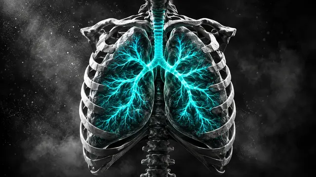
Chest CT read: nodules with Fleischner or Lung-RADS, PE with RV strain, lungs, pleura and ribs.
Chest16 sections
CT· volume
Planning CT contouring: site-specific organs at risk and a GTV proposal as a DICOM RTSTRUCT.
This model is not available to run yet. Vote to show interest and get an email when it opens.

Radiotherapy Planning CT contours the simulation CT before a treatment plan is made. Upload the planning CT series as it leaves the CT simulator, couch, immobilisation mask and all; the run recognises the treatment site (head and neck, thorax, abdomen or pelvis) and contours the full organ-at-risk set for that site.
The contours come back as a DICOM RT Structure Set that a treatment planning system can import, with structure names following AAPM TG-263, organ volumes and contour quality-assurance flags. For lung and for nasopharyngeal and other head and neck cancers the run also proposes an editable gross tumour volume (GTV).
Contouring support for the radiotherapy team. Organ-at-risk delineation is repetitive manual work that delays planning; the run returns the full organ set as editable contours, ready to import into the planning system.
Radiation oncologists, dosimetrists, medical physicists and radiotherapy departments. Developers connect the run to the department's DICOM node so that structure sets arrive in the planning system alongside the simulation CT.
| Requirement | Why |
|---|---|
| Frame of Reference UID | The RT Structure Set must reference the planning CT geometry exactly |
| Image Position and Orientation (Patient) and Pixel Spacing | Contours are written in patient coordinates on the original slices |
| Field of view covering the body outline | A truncated field of view affects body and skin contours and is flagged |
| Treatment site within head and neck, thorax, abdomen or pelvis | The organ set is chosen per site |
Dental metal and implant streaks are reported as quality flags on the structures they cross.
The treatment site and the tumour type can be stated in clinical_context; otherwise the site is detected from the
anatomy in the scan.
A JSON result with structure volumes and QA flags, and the RTSTRUCT that carries the contours.
| Section | Content |
|---|---|
| Organs at risk | Site-specific OAR contours for head and neck, thorax, abdomen or pelvis, with volumes and QA flags |
| GTV proposal | Editable gross tumour volume for lung and nasopharyngeal or head and neck cancer |
| Draft report | Opt-in (options.report): an English narrative that interprets the result for the reader (findings, impression, limitations); every number is checked against the findings |
Every structure carries contour QA flags. Structures near dental metal, immobilisation devices or the edge of a truncated field of view are marked.
One run returns every section its input supports.
Chest CT
Chest CT read: nodules with Fleischner or Lung-RADS, PE with RV strain, lungs, pleura and ribs.
Head CT
Head CT read: bleed volumes, ASPECTS, vessel occlusion, perfusion core and skull fractures.
Abdomen & Pelvis CT
Abdomen-pelvis CT read: acute abdomen flags, organ lesions with management, stones and trauma.
Cardiac CT
Cardiac CT read: Agatston calcium, CAD-RADS 2.0 stenosis, plaque, chambers and TAVI measurements.
Spine CT
Spine CT read: every vertebra labelled, fractures with AO Spine type, alignment and canal size.
CT Angiography (Aorta, Carotid, Run-off)
Arterial CTA read: aortic diameters, dissection and aneurysm, endoleak, carotid and leg stenoses.