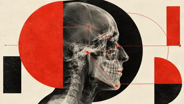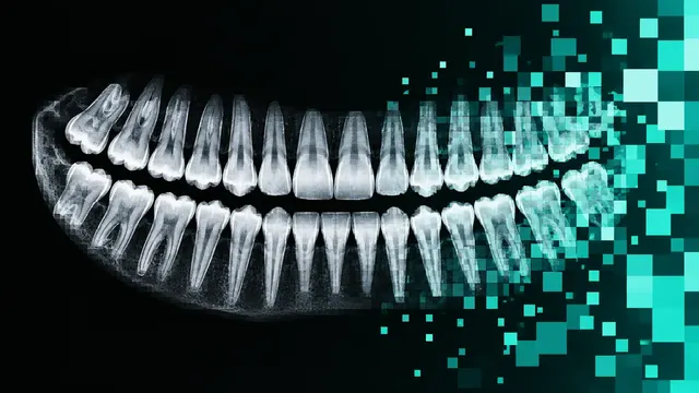
Panoramic Radiograph (OPG)
OPG read: FDI-numbered odontogram, caries, periapical lesions, third molars and bone level.
Oral7 sections
DX· image-2d
CBCT read: segmented teeth, jaws and nerve canals, third-molar canal relations and airway size.
This model is not available to run yet. Vote to show interest and get an email when it opens.
Dental CBCT reads the cone-beam CT volumes taken for implants, impacted teeth, endodontic problems, orthodontics, airway and temporomandibular joint questions. Upload the CBCT series; the run segments the anatomy of the whole field of view and returns it both as DICOM segmentations and as STL meshes that planning software can open.
The segmentation covers every tooth with its FDI number and pulp, the maxilla and mandible, both inferior alveolar canals, the maxillary sinuses, the pharynx and any crowns, bridges or implants, giving a 3D odontogram. On top of it the run measures the relation of impacted teeth to the nerve canal and the volume and narrowest point of the pharyngeal airway. The lesion and TMJ sections report periapical lesions, cysts, sinus disease and degenerative condylar signs across the field of view.
Decision support for the dentist or surgeon who often reviews the CBCT alone: canal proximity before a third-molar extraction, and airway measurements that can support a referral for a sleep study. The result states which regions the field of view covers and which sections were assessed in each.
Oral and maxillofacial surgeons, implantologists, endodontists, orthodontists, general dentists who order CBCT and dentomaxillofacial radiologists. Developers import the meshes and measurements into implant-planning, surgical-guide or orthodontic software.
| Requirement | Why |
|---|---|
| Voxel spacing and orientation | Distances to the canal, sinus floor and airway are reported in millimetres |
| Field of view covering the region in question | Structures outside a small field of view are marked as not assessed |
| Both condyles in the volume | Needed for the TMJ section |
| Pharynx in the volume | Needed for the airway section |
CBCT grey values are not reliable Hounsfield units, so density-based values are labelled as relative. Metal streaks from crowns and implants and patient motion are reported as quality flags on the structures they affect.
The reason for the scan (implant site, third-molar extraction, endodontic problem, orthodontic or airway assessment,
TMJ symptoms) goes in clinical_context and orders the guidance; snoring or sleep symptoms put the airway result in
context.
A JSON result with the 3D odontogram and one section per question, plus DICOM SEG and STL exports.
| Section | Content |
|---|---|
| Anatomy segmentation | Teeth with FDI numbers and pulps, maxilla, mandible, inferior alveolar canals, maxillary sinuses, pharynx, crowns, bridges and implants |
| Impacted teeth | Position, root morphology, contact or distance to the inferior alveolar canal, cortical thinning, Winter and Pell & Gregory class |
| Pharyngeal airway | Airway segmentation, volume, minimal cross-sectional area and the level where it occurs |
| Lesions and incidental findings | Periapical lesions with CBCT-PAI, cysts and tumours, sinus disease and other incidental findings; the result states explicitly whether the lesion screen was performed |
| Temporomandibular joint | Condylar morphology and degenerative signs: erosion, osteophyte, flattening, sclerosis |
| Draft report | Opt-in (options.report): an English narrative that interprets the result for the reader (findings, impression, limitations); every number is checked against the findings |
dental/cbct/implant-planning: choose an implant site by FDI position and get bone height and width, distances to
the canal, sinus floor and adjacent roots, cortical thickness and suggested implant dimensions, with Misch density
classes labelled as relative.One run returns every section its input supports.
Panoramic Radiograph (OPG)
OPG read: FDI-numbered odontogram, caries, periapical lesions, third molars and bone level.
Intra-oral Radiographs (Bitewing and Periapical)
Bitewing and periapical read: surface caries depth, bone levels, PAI and restorations on one chart.
Lateral Cephalogram
Ceph tracing: landmarks, cephalometric analyses, skeletal class and CVM growth stage.
Intra-oral 3D Scan
Intra-oral scan processing: FDI-labelled tooth segmentation, missing teeth and dental landmarks.