Dental CBCT
CBCT read: segmented teeth, jaws and nerve canals, third-molar canal relations and airway size.
Oral6 sections
DX· image-2d
Ceph tracing: landmarks, cephalometric analyses, skeletal class and CVM growth stage.
This model is not available to run yet. Vote to show interest and get an email when it opens.
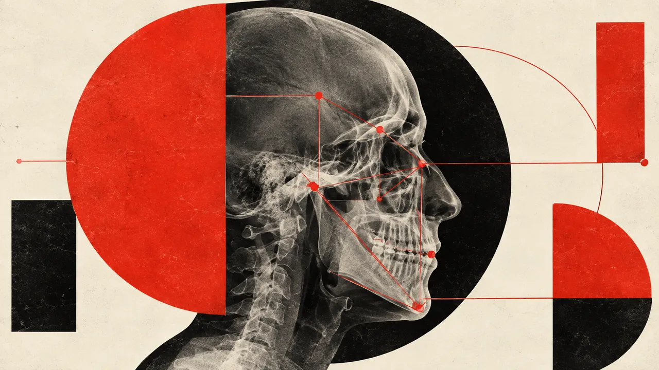
Lateral Cephalogram traces the lateral cephalometric radiograph used for orthodontic and orthognathic planning. Upload one cephalogram; the run calibrates the image scale, places the hard- and soft-tissue landmarks (up to 38) with a confidence for each, and computes the cephalometric analyses from them.
The result gives the skeletal class from ANB and the Wits appraisal, the dental relationships and growth pattern, and the cervical vertebral maturation (CVM) stage read from the second to fourth cervical vertebrae, which tells the orthodontist where the patient is on the growth curve.
Orthodontic decision support: the tracing a clinician would otherwise do by hand, with the same landmark definitions every time, so that measurements are comparable between visits and between clinicians. Growth stage helps time functional treatment. Every value traces to the landmarks it was measured from.
Orthodontists, orthognathic and maxillofacial surgeons, paediatric dentists and orthodontic residents. Developers add automated tracing to orthodontic software, aligner workflows or teaching tools.
| Requirement | Why |
|---|---|
| Scale calibration from the Estimated Radiographic Magnification Factor, pixel spacing or a visible ruler | Linear measurements in millimetres depend on it; without it only angles are reliable |
| C2 to C4 vertebrae in the image | The CVM stage is read from their shape |
| Soft-tissue profile visible | Needed for the soft-tissue landmarks |
| A lateral projection | The landmark set and the analyses are defined on the lateral view |
Bilateral structures such as the mandibular border often project as double contours, and landmarks there carry a lower confidence and a flag. Missing calibration is flagged as well.
Age, sex and the clinical question (growth assessment, orthodontic diagnosis, orthognathic planning) go in
clinical_context. The analyses wanted can be named; otherwise the standard set is returned.
A JSON result with landmark coordinates in millimetres and pixels, the analyses computed from them, and an overlay of the tracing on the original image.
| Section | Content |
|---|---|
| Cephalometric landmarks | Up to 38 hard- and soft-tissue landmarks with coordinates and a confidence per point |
| Cephalometric analyses | Angles and distances for Steiner, Ricketts, McNamara, Downs, Tweed, Wits and Björk–Jarabak; skeletal class I, II or III, dental class and growth pattern |
| CVM stage | Cervical vertebral maturation stage CS1 to CS6 from the C2–C4 vertebrae |
| Draft report | Opt-in (options.report): an English narrative that interprets the result for the reader (findings, impression, limitations); every number is checked against the findings |
dental/ceph/analysis: move landmarks and recompute every analysis, the skeletal class and the growth
pattern from the corrected points.One run returns every section its input supports.
Dental CBCT
CBCT read: segmented teeth, jaws and nerve canals, third-molar canal relations and airway size.
Panoramic Radiograph (OPG)
OPG read: FDI-numbered odontogram, caries, periapical lesions, third molars and bone level.
Intra-oral Radiographs (Bitewing and Periapical)
Bitewing and periapical read: surface caries depth, bone levels, PAI and restorations on one chart.
Intra-oral 3D Scan
Intra-oral scan processing: FDI-labelled tooth segmentation, missing teeth and dental landmarks.