Dental CBCT
CBCT read: segmented teeth, jaws and nerve canals, third-molar canal relations and airway size.
Oral6 sections
DX· image-2d
OPG read: FDI-numbered odontogram, caries, periapical lesions, third molars and bone level.
This model is not available to run yet. Vote to show interest and get an email when it opens.
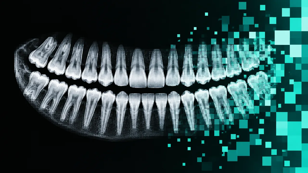
Panoramic Radiograph reads the orthopantomogram (OPG), the screening image taken for new patients and before orthodontic or surgical treatment. Upload one panoramic X-ray; the run finds every tooth, numbers it with the FDI two-digit system, including deciduous and mixed dentitions, and builds an odontogram of the whole mouth.
Each tooth in the odontogram carries its restorative status (fillings, crowns, bridges, root-canal treatment, implants, or missing), caries with depth, periapical radiolucency and an estimated alveolar bone level. Impacted teeth and third molars get their Winter angulation, Pell & Gregory class, eruption level and the radiographic signs of proximity to the inferior alveolar canal. Guidance points to the next image where the panoramic view is not enough, such as a bitewing for early interproximal caries or a CBCT before a high-risk extraction.
Decision support for the dentist who reads the OPG chair-side, usually without a radiologist's report: a complete chart and problem list to check against the clinical examination, and a second look at caries, apical lesions and third-molar risk. Every finding is linked to a tooth number.
General dentists, oral and maxillofacial surgeons, orthodontists, endodontists, periodontists and dental students. Developers bring the odontogram and findings into practice-management software or a dental imaging viewer.
| Requirement | Why |
|---|---|
| A panoramic projection | Bitewings, periapicals and cephalograms are read by their own models |
| Correct patient positioning | Positioning errors distort the teeth and are flagged |
| Left and right orientation | Tooth numbering depends on the side; orientation is normalised before charting |
| Pixel Spacing or Imager Pixel Spacing | Image exports without it carry a calibration warning |
A panoramic image magnifies unevenly across its width, so bone loss is expressed as a percentage of root length and never in millimetres. Ghost images and overlapping premolar contacts are known limits and are flagged where they affect a tooth.
Age (relevant for mixed dentition and third-molar development), the reason for the X-ray and known treatment history
go in clinical_context.
One JSON result with an FDI-numbered odontogram and one section per question; overlays reference the original image.
| Section | Content |
|---|---|
| Tooth numbering | Tooth detection, instance outlines and FDI numbers, including deciduous teeth, and missing teeth |
| Restorative charting | Per tooth: fillings, crowns, bridges, root-canal treatment, implants |
| Caries | Carious lesions per tooth and surface with depth (enamel, dentine, deep or near the pulp) |
| Periapical lesions | Apical radiolucencies (apical periodontitis) per tooth |
| Impacted teeth | Winter angulation, Pell & Gregory class, eruption level and inferior alveolar canal proximity signs |
| Bone loss | Estimated alveolar bone level per tooth and sextant as a percentage of root length, horizontal or vertical pattern |
| Draft report | Opt-in (options.report): an English narrative that interprets the result for the reader (findings, impression, limitations); every number is checked against the findings |
bodySite).One run returns every section its input supports.
Dental CBCT
CBCT read: segmented teeth, jaws and nerve canals, third-molar canal relations and airway size.
Intra-oral Radiographs (Bitewing and Periapical)
Bitewing and periapical read: surface caries depth, bone levels, PAI and restorations on one chart.
Lateral Cephalogram
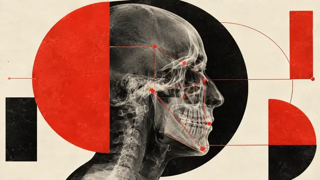
Ceph tracing: landmarks, cephalometric analyses, skeletal class and CVM growth stage.
Intra-oral 3D Scan
Intra-oral scan processing: FDI-labelled tooth segmentation, missing teeth and dental landmarks.