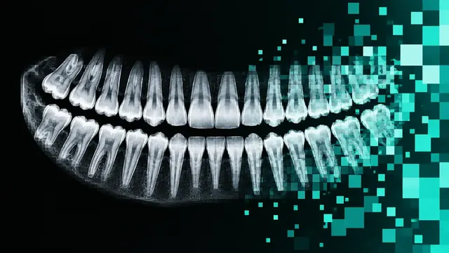
Dental CBCT
CBCT read: segmented teeth, jaws and nerve canals, third-molar canal relations and airway size.
Oral6 sections
DX· image-2d
Intra-oral scan processing: FDI-labelled tooth segmentation, missing teeth and dental landmarks.
This model is not available to run yet. Vote to show interest and get an email when it opens.

Intra-oral 3D Scan processes the triangle meshes produced by intra-oral scanners, the digital impressions behind orthodontic set-ups, clear aligners and restorations. Upload the upper jaw, the lower jaw or both; the run cleans each mesh, aligns it to the occlusal plane and labels every vertex as gingiva or as a numbered tooth.
The result is a segmented dentition with FDI numbers, a list of missing teeth, and dental landmarks on every tooth: cusp tips, mesial and distal contact points and facial axis points. Together they replace the manual step of cutting teeth apart and placing reference points before a set-up or a restoration can be designed.
A processing step for digital dentistry: the clinician, orthodontist or dental technician gets separated, numbered teeth and reference points to start a design from, ready to open in their own planning or CAD software.
Orthodontists, aligner providers, restorative and prosthodontic dentists, dental laboratories and CAD technicians. Developers call the run from aligner-planning, CAD/CAM or model-analysis software to skip manual tooth segmentation.
Each jaw arrives as its own mesh, so every arch is numbered within its own FDI quadrants. Before segmentation, each mesh is cleaned and aligned to the occlusal plane, which puts scans from different scanners into the same orientation for numbering and landmark placement.
The purpose of the scan (orthodontic set-up, aligners, restoration) and known missing or extracted teeth go in
clinical_context, and they are kept with the result.
A JSON result with the per-tooth labels and landmarks, plus the labelled meshes. Tooth labels can be returned in FDI, Universal or Palmer notation, to match the numbering of the receiving software.
| Section | Content |
|---|---|
| Tooth segmentation | Per-vertex segmentation of gingiva and teeth with FDI labels, and the list of missing teeth |
| Dental landmarks | Per tooth: cusp tips, mesial and distal contact points and facial axis points as 3D coordinates |
| Draft report | Opt-in (options.report): an English narrative that interprets the result for the reader (findings, impression, limitations); every number is checked against the findings |
The segmented scan pairs with Dental CBCT for prosthetically driven implant planning: the crowns come from the intra-oral scan, and bone and nerve canal from the CBCT and its implant-planning tool.
One run returns every section its input supports.
Dental CBCT
CBCT read: segmented teeth, jaws and nerve canals, third-molar canal relations and airway size.
Panoramic Radiograph (OPG)
OPG read: FDI-numbered odontogram, caries, periapical lesions, third molars and bone level.
Intra-oral Radiographs (Bitewing and Periapical)
Bitewing and periapical read: surface caries depth, bone levels, PAI and restorations on one chart.
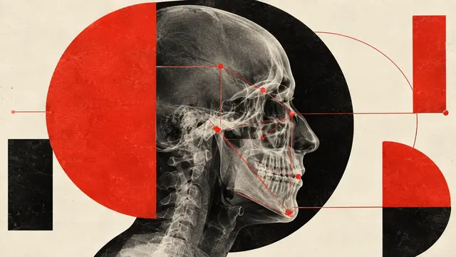
Lateral Cephalogram
Ceph tracing: landmarks, cephalometric analyses, skeletal class and CVM growth stage.