Brain MRI
Brain MRI read: stroke, tumours, MS lesions, aneurysms, atrophy and ARIA in one result.
Neuro15 sections
MR· volume
Breast MRI read: enhancing lesions, kinetics, BI-RADS assessment, BPE, FGT and implant integrity.
This model is not available to run yet. Vote to show interest and get an email when it opens.

Breast MRI reads dynamic contrast-enhanced studies for high-risk screening, preoperative staging, problem solving, monitoring of neoadjuvant chemotherapy and implant assessment. The run orders the dynamic phases by acquisition time, motion-corrects them, computes subtractions and kinetic curves, and reads the breasts the way the BI-RADS MRI lexicon is structured: composition first, then enhancement, then findings.
It grades the amount of fibroglandular tissue (a–d) and the background parenchymal enhancement with its symmetry, detects and segments mass and non-mass enhancement with size and kinetic curve type, and assigns BI-RADS descriptors and a final assessment category 0–6. On a baseline and on-treatment pair, it reports functional tumour volume change and a pCR likelihood; on silicone-sensitive sequences it looks for intracapsular and extracapsular rupture.
Decision support for physicians reporting breast MRI: lesion detection across hundreds of dynamic images, kinetic curves that would otherwise be drawn by hand, and a BI-RADS assessment set in the context of FGT and BPE. Background enhancement varies with the menstrual cycle and therapy, which is why both can be passed as context.
Breast imagers and general radiologists covering supplemental screening of dense breasts, breast surgeons planning resection, medical oncologists following neoadjuvant therapy, and plastic surgeons assessing implants. Developers call the run to bring BI-RADS-structured MRI results into screening registries and breast-centre software.
prior_files for comparison and for neoadjuvant response.TemporalPositionIdentifier or acquisition time.| Requirement | Why |
|---|---|
| Non-contrast T1 | Fibroglandular tissue segmentation and FGT category |
| Pre-contrast and at least two post-contrast T1 phases | Lesion detection, initial enhancement and delayed curve type |
| Subtractions (computed when absent) | Lesion conspicuity and BPE grading |
| T2 (optional) | Lesion characterisation, cysts and lymph nodes |
| DWI (optional) | Additional characterisation and treatment response |
| Silicone-sensitive sequences (implant studies) | Rupture signs |
Indication (screening, staging, problem solving, neoadjuvant monitoring, implant), risk status, menstrual phase or
hormonal therapy, and treatment dates go in clinical_context.
| Section | Content |
|---|---|
| Fibroglandular tissue | Breast and FGT masks, FGT %, category a–d |
| Background parenchymal enhancement | BPE level (minimal, mild, moderate, marked) and symmetry |
| Lesions and kinetics | Mass and non-mass enhancement: 3D mask, size, location and kinetic curve type |
| BI-RADS assessment | BI-RADS lexicon descriptors per lesion and the final assessment category 0–6 |
| Neoadjuvant therapy response | Functional tumour volume change from baseline and pCR likelihood |
| Implant integrity | Intracapsular and extracapsular rupture signs |
| Draft report | Opt-in (options.report): an English narrative that interprets the result for the reader (findings, impression, limitations); every number is checked against the findings |
The result presents FGT and BPE before the lesions so the assessment is read in context: moderate, marked or asymmetric BPE can mask or mimic lesions.
One run returns every section its input supports.
Brain MRI
Brain MRI read: stroke, tumours, MS lesions, aneurysms, atrophy and ARIA in one result.
Prostate MRI
Prostate MRI read: PI-RADS v2.1 lesions, sector map, PSA density and fusion-biopsy targets.
Spine MRI
Spine MRI read: a graded level table, cord measurements and red flags in Fardon 2014 terms.
Abdominal MRI
Abdominal MRI read: liver fat and iron, LI-RADS, MRCP, pancreatic cysts, Bosniak and kidney volume.
Cardiac MRI
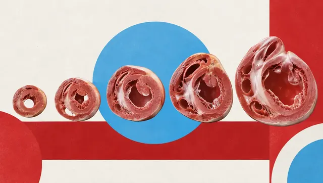
Cardiac MRI read: ventricular function, scar, T1/T2/ECV mapping, flow and strain per AHA segment.
Knee MRI
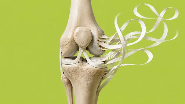
Knee MRI read: ACL status, meniscal tears, cartilage defects and bone marrow oedema.