Brain MRI
Brain MRI read: stroke, tumours, MS lesions, aneurysms, atrophy and ARIA in one result.
Neuro15 sections
MR· volume
Prostate MRI read: PI-RADS v2.1 lesions, sector map, PSA density and fusion-biopsy targets.
This model is not available to run yet. Vote to show interest and get an email when it opens.

Prostate MRI reads biparametric (T2W and diffusion) and multiparametric (with DCE) studies in the MRI-first diagnostic pathway. The run segments the gland and its peripheral and transition zones, measures the volume and, with a PSA value, the PSA density. It then detects suspicious lesions, segments each one, places it on the PI-RADS v2.1 sector map and assigns a PI-RADS category.
Around the lesion list, the result states whether each sequence is diagnostically adequate (PI-QUAL), flags extraprostatic extension and seminal vesicle invasion per side, and exports biopsy targets that fusion-biopsy systems import. With a prior study on active surveillance, lesions are matched and scored with PRECISE; after prostatectomy or radiotherapy, the read follows PI-RR for local recurrence.
Decision support for physicians reporting prostate MRI, where transition-zone lesions and PI-RADS 3 versus 4 calls vary most between readers: a consistent lesion list with measurements and sector locations, and a quality statement that says when image quality limits a negative read.
Radiologists who report prostate MRI at any volume, urologists planning targeted biopsy or active surveillance, and radiation oncologists. Through the API, targets and contours flow to fusion-biopsy platforms and urology registries.
prior_files for PRECISE assessment on active surveillance.| Requirement | Why |
|---|---|
| Axial T2W | Reference series for zones, lesion shape and transition-zone scoring |
| DWI with a high b-value (≥1400 s/mm²) | Dominant sequence for the peripheral zone; computed from the acquired b-values when absent |
| ADC map | Scores diffusion restriction with the high-b images; computed from DWI when not supplied |
| DCE (optional) | A positive DCE upgrades a peripheral-zone DWI score of 3 to PI-RADS 4; also supports local staging and recurrence reads |
Missing or degraded DWI (rectal gas, hip prostheses) and post-biopsy haemorrhage are reported in the quality section rather than silently scored.
PSA (for PSA density), prior biopsy results, treatment history (prostatectomy, radiotherapy) and active-surveillance
status go in clinical_context.
| Section | Content |
|---|---|
| Image quality (PI-QUAL) | Per-sequence diagnostic adequacy, artefacts (gas, motion, distortion) and the PI-QUAL score |
| Gland and zones | Whole-gland, peripheral-zone and transition-zone masks, gland volume, PSA density when PSA is given |
| PI-RADS lesions | Each lesion with mask, size, zone, sector-map location, PI-RADS v2.1 category and a biopsy target |
| Local staging | Likelihood of extraprostatic extension, seminal vesicle and neurovascular-bundle invasion per side |
| Active surveillance (PRECISE) | Lesion matching against the prior, size change and PRECISE score |
| Recurrence (PI-RR) | Local recurrence likelihood and location after prostatectomy or radiotherapy |
| Draft report | Opt-in (options.report): an English narrative that interprets the result for the reader (findings, impression, limitations); every number is checked against the findings |
sector_map block), with the PI-RADS v2.1
features for extraprostatic extension.Gland volume comes from the segmentation in millilitres, so PSA density is PSA divided by that volume. The PI-RADS category follows the dominant sequence of each zone: diffusion in the peripheral zone, T2W in the transition zone.
One run returns every section its input supports.
Brain MRI
Brain MRI read: stroke, tumours, MS lesions, aneurysms, atrophy and ARIA in one result.
Spine MRI
Spine MRI read: a graded level table, cord measurements and red flags in Fardon 2014 terms.
Abdominal MRI
Abdominal MRI read: liver fat and iron, LI-RADS, MRCP, pancreatic cysts, Bosniak and kidney volume.
Breast MRI
Breast MRI read: enhancing lesions, kinetics, BI-RADS assessment, BPE, FGT and implant integrity.
Cardiac MRI
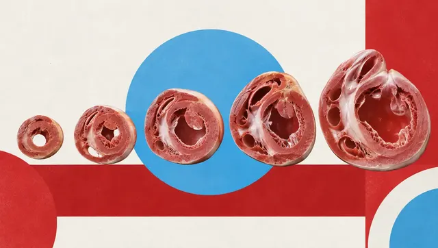
Cardiac MRI read: ventricular function, scar, T1/T2/ECV mapping, flow and strain per AHA segment.
Knee MRI
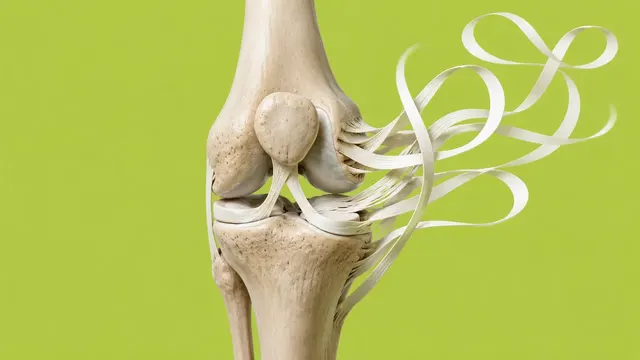
Knee MRI read: ACL status, meniscal tears, cartilage defects and bone marrow oedema.