Brain MRI
Brain MRI read: stroke, tumours, MS lesions, aneurysms, atrophy and ARIA in one result.
Neuro15 sections
MR· volume
Cardiac MRI read: ventricular function, scar, T1/T2/ECV mapping, flow and strain per AHA segment.
This model is not available to run yet. Vote to show interest and get an email when it opens.
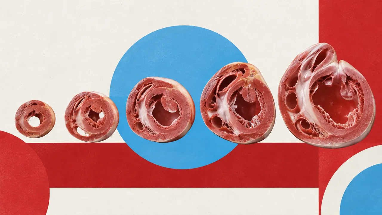
Cardiac MRI reads a complete CMR study: cine short-axis and long-axis stacks, late gadolinium enhancement and, when acquired, parametric maps, phase-contrast flow and first-pass perfusion. The run contours the left ventricle, myocardium and right ventricle on every cine phase, detects end-diastole and end-systole from the volume curve, and computes volumes, ejection fraction and mass indexed to body surface area.
Tissue characterisation is reported on the AHA 17-segment model: scar extent and transmurality on LGE with an ischaemic or non-ischaemic pattern, segmental native T1, T2 and extracellular volume, and perfusion defects. Flow, shunt ratio and strain complete the study, and the sections combine into a pattern-based differential, including the Lake Louise 2018 criteria for myocarditis.
Decision support for physicians reporting CMR, where manual contouring is slow and basal-slice and papillary-muscle choices change EF and mass: reproducible volumes, segmental tissue values and a viability map after myocardial infarction.
Cardiologists and radiologists who report CMR, imaging fellows, heart-failure and cardiomyopathy clinics, and interventional teams who need segmental transmurality before revascularising a stenosed territory. Developers call the run to add CMR quantification to a cardiovascular PACS or registry.
TriggerTime and CardiacNumberOfImages.| Requirement | Why |
|---|---|
| Short-axis cine stack covering both ventricles | Volumes, EF and mass |
| 2-, 3- and 4-chamber cine (optional) | Atria, long-axis function and strain |
| LGE short-axis | Scar, transmurality and microvascular obstruction per segment |
| Native T1 and T2 maps (optional) | Segmental T1 and T2; post-contrast T1 with a haematocrit adds ECV |
| 2D phase contrast with a valid VENC (optional) | Flow volumes, regurgitant fraction, Qp:Qs; aliasing is checked |
| Stress and rest first-pass perfusion (optional) | Segmental perfusion defects |
Height and weight index volumes and mass to body surface area. A same-day haematocrit is needed for ECV. The indication (chest pain, suspected myocarditis, cardiomyopathy, viability) shapes the differential. T1 and T2 values are compared with local normal ranges, because mapping values are scanner- and sequence-specific.
| Section | Content |
|---|---|
| Chamber segmentation | LV, myocardium and RV masks on every cine phase, plus atria on long-axis views |
| Ventricular function | LV and RV EDV, ESV, SV and EF, LV mass, indexed to BSA and flagged against reference ranges |
| Late gadolinium enhancement | Scar mask, scar as % of LV mass, transmurality per segment, ischaemic or non-ischaemic pattern, microvascular obstruction |
| T1, T2 and ECV mapping | Myocardial masks on the maps, per-segment native T1, T2 and ECV against local normal ranges |
| Stress perfusion | Perfusion defects per AHA segment; myocardial blood flow when vendor quantitative maps are supplied |
| Phase-contrast flow | Vessel contour, forward and backward volume, regurgitant fraction, peak velocity, Qp:Qs |
| Myocardial strain | Global and segmental longitudinal, circumferential and radial strain |
| Draft report | Opt-in (options.report): an English narrative that interprets the result for the reader (findings, impression, limitations); every number is checked against the findings |
aha_segments block.Volumes and mass are reported in absolute terms and, with height and weight, indexed to BSA. Quantitative maps are kept in physical units (ms for T1 and T2, % for ECV) and are never intensity-normalised.
One run returns every section its input supports.
Brain MRI
Brain MRI read: stroke, tumours, MS lesions, aneurysms, atrophy and ARIA in one result.
Prostate MRI
Prostate MRI read: PI-RADS v2.1 lesions, sector map, PSA density and fusion-biopsy targets.
Spine MRI
Spine MRI read: a graded level table, cord measurements and red flags in Fardon 2014 terms.
Abdominal MRI
Abdominal MRI read: liver fat and iron, LI-RADS, MRCP, pancreatic cysts, Bosniak and kidney volume.
Breast MRI
Breast MRI read: enhancing lesions, kinetics, BI-RADS assessment, BPE, FGT and implant integrity.
Knee MRI
Knee MRI read: ACL status, meniscal tears, cartilage defects and bone marrow oedema.