Brain MRI
Brain MRI read: stroke, tumours, MS lesions, aneurysms, atrophy and ARIA in one result.
Neuro15 sections
MR· volume
Knee MRI read: ACL status, meniscal tears, cartilage defects and bone marrow oedema.
This model is not available to run yet. Vote to show interest and get an email when it opens.
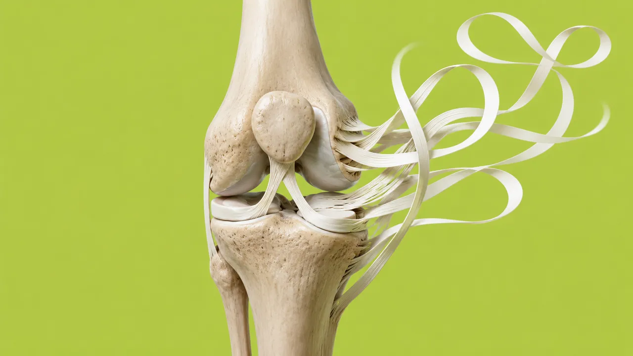
Knee MRI reads the highest-volume joint MRI, ordered for twisting injuries, locking, effusion and suspected osteoarthritis. The run takes the standard multiplanar protocol and works structure by structure, the way a musculoskeletal radiologist reads a knee: ligament, menisci, articular cartilage and subchondral bone.
It classifies the anterior cruciate ligament as intact, partially torn or completely torn with its secondary signs, locates meniscal tears by meniscus and horn and measures extrusion, segments the cartilage plates into a thickness map with focal defects, and describes bone marrow oedema and contusion patterns with lesion size. The result is organised one structure per section.
Decision support for physicians reporting knee MRI: a structured checklist so that a meniscal root or posterior-horn tear is not overlooked on a busy list, and cartilage measurements that would take too long by hand. Every finding is shown on the image it was read from.
General and musculoskeletal radiologists and teleradiologists reading knee MRI, and orthopaedic and sports-medicine surgeons deciding between rehabilitation, arthroscopy and reconstruction. Developers use the cartilage segmentation and thickness maps for osteoarthritis research and longitudinal follow-up.
| Requirement | Why |
|---|---|
| Sagittal and coronal PD or T2 fat-sat | ACL fibres, meniscal horns and body |
| Axial fat-sat sequence | Patellofemoral cartilage and a third plane for meniscal tears |
| Fluid-sensitive fat-sat sequences | Bone marrow oedema and contusion pattern |
| T1 (optional) | Marrow reference and fracture lines |
A study without fat-suppressed fluid-sensitive images cannot be graded for marrow oedema; the result says so instead of returning an empty section.
Mechanism of injury, the side, prior surgery (ACL reconstruction, meniscectomy) and the clinical question go in
clinical_context. Prior surgery matters: graft and repaired-meniscus appearances are read differently from native
tissue.
| Section | Content |
|---|---|
| Anterior cruciate ligament | Intact, partial tear or complete tear, with secondary signs |
| Menisci | Tear presence, medial or lateral meniscus, horn and location, extrusion |
| Cartilage | Cartilage segmentation, thickness map, focal defects with an ICRS or MOAKS-style grade |
| Bone marrow oedema | Bone marrow oedema and contusion pattern, bone marrow lesion size with a MOAKS-style grade |
| Draft report | Opt-in (options.report): an English narrative that interprets the result for the reader (findings, impression, limitations); every number is checked against the findings |
The ACL and meniscal sections answer the acute-injury question; cartilage and bone marrow lesions answer the degenerative one. A pivot-shift contusion pattern (lateral femoral condyle and posterolateral tibial plateau) is a secondary sign of an ACL tear, so the two sections are read together.
One run returns every section its input supports.
Brain MRI
Brain MRI read: stroke, tumours, MS lesions, aneurysms, atrophy and ARIA in one result.
Prostate MRI
Prostate MRI read: PI-RADS v2.1 lesions, sector map, PSA density and fusion-biopsy targets.
Spine MRI
Spine MRI read: a graded level table, cord measurements and red flags in Fardon 2014 terms.
Abdominal MRI
Abdominal MRI read: liver fat and iron, LI-RADS, MRCP, pancreatic cysts, Bosniak and kidney volume.
Breast MRI
Breast MRI read: enhancing lesions, kinetics, BI-RADS assessment, BPE, FGT and implant integrity.
Cardiac MRI
Cardiac MRI read: ventricular function, scar, T1/T2/ECV mapping, flow and strain per AHA segment.