Brain MRI
Brain MRI read: stroke, tumours, MS lesions, aneurysms, atrophy and ARIA in one result.
Neuro15 sections
MR· volume
Fetal brain MRI read: 3D reconstruction, tissue volumes and biometry against gestational age.
This model is not available to run yet. Vote to show interest and get an email when it opens.
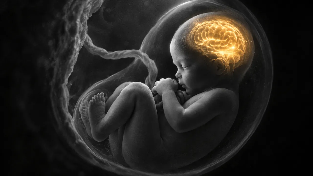
Fetal MRI reads the fetal brain examination performed after a suspicious anomaly scan, above all for ventriculomegaly and other central nervous system malformations. The fetus moves between and during slices, so the stacks of single-shot T2 images acquired in different planes are each motion-corrupted and incomplete on their own. The run first combines them by slice-to-volume reconstruction into one motion-corrected, high-resolution 3D brain volume.
On that volume it segments the brain tissues and reports their volumes, and it measures the standard fetal brain biometry: biparietal and occipitofrontal diameters, transcerebellar diameter and the atrial width of the lateral ventricles. Each measurement is placed against gestational-age reference values, so a borderline atrial width or a small cerebellum is reported with how far it sits from the expected range.
Decision support for physicians reading fetal MRI in specialised centres and for those who see the exam only occasionally: reproducible biometry taken on a reconstructed volume rather than on oblique slices, and tissue volumes that cannot be measured by hand in clinical time. Measurements are shown on the reconstructed images.
Paediatric and fetal neuroradiologists, general radiologists covering prenatal imaging, maternal-fetal medicine specialists and fetal neurologists who counsel families after an abnormal scan. Researchers use the reconstruction and tissue volumes for fetal brain development studies.
| Requirement | Why |
|---|---|
| Several single-shot T2 stacks of the fetal brain in different orientations | Slice-to-volume reconstruction needs overlapping views to correct motion |
Gestational age in clinical_context |
Biometry and volumes are interpreted against age-specific references |
Motion between stacks is expected and corrected; a study whose stacks cannot be reconstructed into a usable volume returns a quality block instead of measurements.
Besides gestational age, the anomaly-scan finding that prompted the MRI (for example ventriculomegaly measured on
ultrasound) goes in clinical_context.
| Section | Content |
|---|---|
| Brain tissue segmentation | Tissue labels on the reconstructed volume with per-tissue volumes |
| Brain biometry | Biparietal and occipitofrontal diameters, transcerebellar diameter and atrial width against gestational-age references |
| Draft report | Opt-in (options.report): an English narrative that interprets the result for the reader (findings, impression, limitations); every number is checked against the findings |
Diameters measured on a single motion-corrupted slice depend on how the slice happens to cut the head. On a reconstructed volume, the diameters are measured in planes aligned to the fetal brain itself, so they depend less on slice angle and can be compared between examinations.
One run returns every section its input supports.
Brain MRI
Brain MRI read: stroke, tumours, MS lesions, aneurysms, atrophy and ARIA in one result.
Prostate MRI
Prostate MRI read: PI-RADS v2.1 lesions, sector map, PSA density and fusion-biopsy targets.
Spine MRI
Spine MRI read: a graded level table, cord measurements and red flags in Fardon 2014 terms.
Abdominal MRI
Abdominal MRI read: liver fat and iron, LI-RADS, MRCP, pancreatic cysts, Bosniak and kidney volume.
Breast MRI
Breast MRI read: enhancing lesions, kinetics, BI-RADS assessment, BPE, FGT and implant integrity.
Cardiac MRI
Cardiac MRI read: ventricular function, scar, T1/T2/ECV mapping, flow and strain per AHA segment.