Brain MRI
Brain MRI read: stroke, tumours, MS lesions, aneurysms, atrophy and ARIA in one result.
Neuro15 sections
MR· volume
Pelvic MRI read: rectal cancer staging, gynaecological staging, O-RADS MRI, fibroids and fistulas.
This model is not available to run yet. Vote to show interest and get an email when it opens.
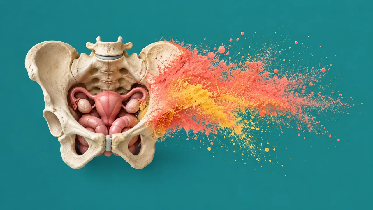
Pelvic MRI reads the non-prostate pelvic protocols: rectal cancer staging and restaging, cervical and endometrial cancer, adnexal masses that are indeterminate on ultrasound, fibroids and adenomyosis, endometriosis and perianal fistula. Each indication has its own structured checklist, so the run first recognises the protocol (for example an oblique high-resolution T2 angled to the rectal tumour) and then returns the section that matches it.
For rectal cancer it reports the full staging checklist: tumour height, T stage, distance to the mesorectal fascia, extramural venous invasion, nodes and, after neoadjuvant therapy, the MR tumour regression grade. Fascia involvement and venous invasion are the items generalists most often miss. For gynaecological cancers it returns the FIGO stage with the invasion features behind it; for adnexal masses, the O-RADS MRI score; for benign disease, a fibroid map with FIGO types, an endometriosis map and a fistula classification.
Decision support for physicians reporting pelvic MRI against checklists that are long and indication-specific: a complete structured read for the multidisciplinary meeting, with each stage-defining feature stated and located. Every staging item comes with its image evidence.
Abdominal, pelvic and general radiologists; colorectal surgeons and oncologists in rectal cancer pathways; gynaecological oncologists; gynaecologists treating fibroids, adenomyosis and endometriosis; and gastroenterologists and surgeons managing perianal Crohn's disease.
prior_files for rectal restaging after neoadjuvant treatment.| Section | Needs |
|---|---|
| Rectal cancer staging | High-resolution oblique axial T2, ± DWI |
| Cervical cancer staging | T2 and DWI |
| Endometrial cancer staging | T2, DWI and DCE |
| Adnexal mass (O-RADS MRI) | T2, DWI and DCE |
| Fibroids and adenomyosis | Sagittal and axial T2, ± post-contrast |
| Endometriosis | T2 and fat-suppressed T1 |
| Perianal fistula | Fat-suppressed T2 and post-contrast T1 |
The indication, treatment status (before or after chemoradiotherapy), menopausal status and the clinical question go in
clinical_context; they select the checklist when the protocol alone is ambiguous.
| Section | Content |
|---|---|
| Rectal cancer staging | Tumour height, T stage, MRF/CRM distance, EMVI, nodes; mrTRG on restaging |
| Cervical cancer staging | Tumour size, parametrial and vaginal invasion, nodes, FIGO stage |
| Endometrial cancer staging | Depth of myometrial invasion, cervical stromal invasion, FIGO stage |
| Adnexal mass (O-RADS MRI) | Adnexal mass characterisation and O-RADS MRI score 1–5 |
| Fibroids and adenomyosis | Fibroid count, size and FIGO type, uterine volume, adenomyosis flag |
| Endometriosis | Endometriomas, ovary segmentation, map of deep infiltrating endometriosis |
| Perianal fistula | Fistula tract classification, abscess and activity |
| Draft report | Opt-in (options.report): an English narrative that interprets the result for the reader (findings, impression, limitations); every number is checked against the findings |
figo_stage block.One run returns every section its input supports.
Brain MRI
Brain MRI read: stroke, tumours, MS lesions, aneurysms, atrophy and ARIA in one result.
Prostate MRI
Prostate MRI read: PI-RADS v2.1 lesions, sector map, PSA density and fusion-biopsy targets.
Spine MRI
Spine MRI read: a graded level table, cord measurements and red flags in Fardon 2014 terms.
Abdominal MRI
Abdominal MRI read: liver fat and iron, LI-RADS, MRCP, pancreatic cysts, Bosniak and kidney volume.
Breast MRI
Breast MRI read: enhancing lesions, kinetics, BI-RADS assessment, BPE, FGT and implant integrity.
Cardiac MRI
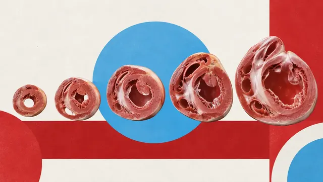
Cardiac MRI read: ventricular function, scar, T1/T2/ECV mapping, flow and strain per AHA segment.