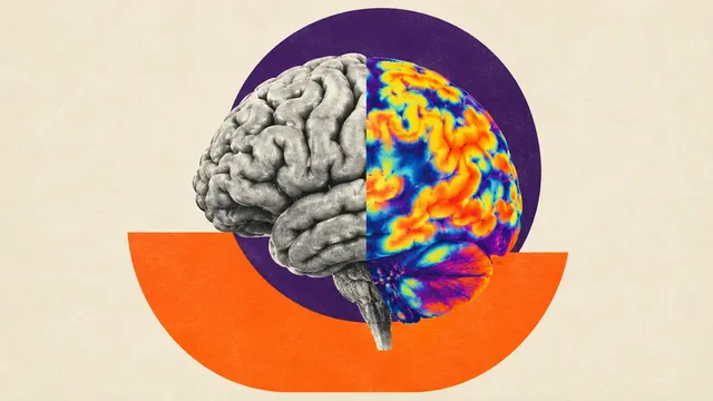
Brain PET and DaT SPECT
Brain PET and DaT SPECT read: Centiloid, CenTauRz, FDG dementia patterns and striatal binding.
Neuro6 sections
NM· volume
Lung V/Q read: segmental mismatched perfusion defects and a pulmonary embolism call per EANM 2019.
This model is not available to run yet. Vote to show interest and get an email when it opens.
Lung V/Q Scintigraphy reads the ventilation/perfusion study, the preferred test for pulmonary embolism when CT pulmonary angiography is a poor choice: in pregnancy, in renal failure and in patients with contrast allergy. It is also the screening test for chronic thromboembolic disease before a CTEPH work-up. Upload the ventilation and perfusion images, planar or tomographic, with the CT of a SPECT/CT when there is one.
The run compares the two acquisitions segment by segment. A perfusion defect in a region that is normally ventilated is a mismatch, the scintigraphic sign of embolism; matched defects point to parenchymal or airway disease instead. The result lists the mismatched defects by lung segment and gives the overall call that the EANM guideline asks for: PE present, PE absent or non-diagnostic.
Decision support for a study that emergency physicians often need out of hours, when an experienced reader may not be on site: a segment-level map of mismatched defects and a guideline-structured conclusion. In suspected chronic thromboembolic disease, the defect map documents the extent of residual perfusion loss.
Nuclear-medicine physicians and radiologists who report V/Q studies, emergency physicians, obstetric teams and pulmonologists who act on the result, and pulmonary hypertension services screening for CTEPH.
| Requirement | Why |
|---|---|
| Ventilation and perfusion acquisitions of the same examination | Mismatch is defined by comparing the two |
| Matching projections or a tomographic acquisition for both | Defects are compared region by region |
| Original DICOM NM images, not screenshots | Count distribution and segmental mapping need the source data |
| CT of a SPECT/CT (optional) | Anatomical context for matched defects, such as consolidation or effusion |
Tomographic V/Q, the acquisition the EANM 2019 guideline recommends, allows segmental mapping in three dimensions; planar studies are read by projection.
Pregnancy, the question asked (acute embolism or chronic thromboembolic disease), recent chest radiograph or CT
findings and known lung disease can be passed in clinical_context; they shape the guidance.
| Section | Content |
|---|---|
| Pulmonary embolism | Mismatched perfusion defects by lung segment, matched defects, and an overall call: PE present, PE absent or non-diagnostic |
| Draft report | Opt-in (options.report): an English narrative that interprets the result for the reader (findings, impression, limitations); every number is checked against the findings |
The defects are shown on the ventilation and perfusion images side by side, so each mismatch can be checked against the regions it was drawn from.
Embolism blocks blood flow while the airways stay open, so perfusion falls in a region whose ventilation is normal. Counting mismatched defects at the level of lung segments and subsegments is how the EANM criteria separate embolism from the matched defects of pneumonia, emphysema or airway disease.
One run returns every section its input supports.
Brain PET and DaT SPECT
Brain PET and DaT SPECT read: Centiloid, CenTauRz, FDG dementia patterns and striatal binding.
FDG PET/CT (Oncology)
Oncologic FDG PET/CT read: lesions with SUV, Lugano stage, Deauville score and metabolic response.
Bone Scintigraphy
Whole-body bone scan read: metastatic hotspots, Bone Scan Index, PCWG3 progression and ATTR-CM flag.
Nuclear Cardiology (MPI and Cardiac Amyloid)
Nuclear cardiology read: perfusion scores, gated function, myocardial blood flow and ATTR-CM grade.
PSMA PET/CT
PSMA PET/CT read: PROMISE V2 miTNM staging, PSMA tumour volume and Lu-PSMA eligibility signals.
Renal Scintigraphy
Renal scintigraphy read: diuretic renogram curves, split function, drainage and DMSA cortical scars.