FDG PET/CT (Oncology)
Oncologic FDG PET/CT read: lesions with SUV, Lugano stage, Deauville score and metabolic response.
Whole body9 sections
NM· volume
Brain PET and DaT SPECT read: Centiloid, CenTauRz, FDG dementia patterns and striatal binding.
This model is not available to run yet. Vote to show interest and get an email when it opens.
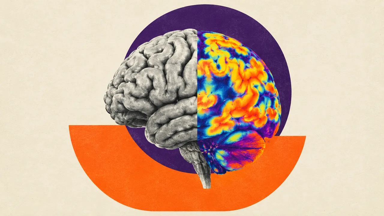
Brain PET and DaT SPECT reads the four neuro-nuclear exams that memory and movement-disorder clinics order: amyloid PET, tau PET, FDG brain PET and dopamine transporter (DaT) SPECT. Upload the reconstructed brain study; the tracer is identified from the DICOM radiopharmaceutical codes, the scan is spatially normalised (an MRI is optional) and the quantification that belongs to that tracer is returned.
Amyloid PET gets a Centiloid value, SUVR and a positive or negative classification with visual-read support. Tau PET gets CenTauRz, regional SUVR and a Braak-like stage. FDG PET gets regional z-score maps against a normal database and the probabilities of Alzheimer's, frontotemporal and Lewy-body patterns. DaT SPECT gets caudate and putamen striatal binding ratios with asymmetry. With a prior scan, the run reports the change, such as the fall in Centiloid during anti-amyloid therapy.
Decision support where a number is part of the clinical question: amyloid status before anti-amyloid antibodies, amyloid clearance on treatment, an aetiological pattern in cognitive decline, presynaptic dopaminergic loss in parkinsonism. Atrophy, partial-volume effects, off-target tau binding, head motion and vascular lesions on DaT SPECT all affect the numbers, and the result shows the visual-read support beside each value.
Nuclear-medicine physicians and neuroradiologists who read brain PET and DaT SPECT, neurologists in memory and movement-disorder clinics who ask for a Centiloid or binding ratio, and research groups running longitudinal amyloid or tau cohorts.
prior_files): an earlier scan with the same tracer for the change section.| Requirement | Why |
|---|---|
| Reconstructed tomographic data, not screenshots or summary pages | Spatial normalisation and regional quantification need the volume |
| Radiopharmaceutical code in the DICOM header | Selects tracer-specific reference regions and thresholds |
| Whole brain in the field of view | Tracer-specific reference regions for SUVR and binding ratios |
| Limited head motion | Motion blurs cortical and striatal uptake |
Reference regions and positivity thresholds are tracer-specific, and the normal database used for z-scores is named with its version in the result.
Age, sex, the clinical question (cognitive impairment, parkinsonism) and current anti-amyloid treatment can be passed
in clinical_context; they shape the guidance, not the measured values.
| Section | Content |
|---|---|
| Amyloid PET (Centiloid) | Centiloid value, SUVR, amyloid positive or negative with visual-read support |
| Tau PET (CenTauRz) | CenTauRz, regional SUVR, Braak-like stage and visual-read pattern |
| FDG dementia patterns | Regional z-score maps against a normal database; Alzheimer's, frontotemporal and Lewy-body pattern probabilities |
| DaT SPECT striatal binding | Caudate and putamen striatal binding ratios, asymmetry and normal or abnormal probability |
| Change versus prior | Change in Centiloid, CenTauRz or FDG z-scores against the prior scan |
| Draft report | Opt-in (options.report): an English narrative that interprets the result for the reader (findings, impression, limitations); every number is checked against the findings |
The sections that appear depend on the tracer: an amyloid study returns the amyloid section, and the change section appears only with a prior.
Centiloid maps amyloid PET from different tracers and pipelines onto one scale anchored at 0 for young controls and 100 for typical Alzheimer's disease. That shared scale makes the change section meaningful during anti-amyloid therapy, where amyloid clearance is judged on the Centiloid value over time.
One run returns every section its input supports.
FDG PET/CT (Oncology)
Oncologic FDG PET/CT read: lesions with SUV, Lugano stage, Deauville score and metabolic response.
Bone Scintigraphy
Whole-body bone scan read: metastatic hotspots, Bone Scan Index, PCWG3 progression and ATTR-CM flag.
Nuclear Cardiology (MPI and Cardiac Amyloid)
Nuclear cardiology read: perfusion scores, gated function, myocardial blood flow and ATTR-CM grade.
PSMA PET/CT
PSMA PET/CT read: PROMISE V2 miTNM staging, PSMA tumour volume and Lu-PSMA eligibility signals.
Lung V/Q Scintigraphy
Lung V/Q read: segmental mismatched perfusion defects and a pulmonary embolism call per EANM 2019.
Renal Scintigraphy
Renal scintigraphy read: diuretic renogram curves, split function, drainage and DMSA cortical scars.