Brain PET and DaT SPECT
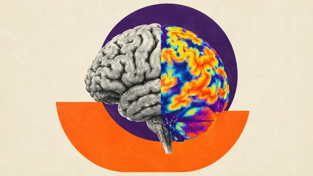
Brain PET and DaT SPECT read: Centiloid, CenTauRz, FDG dementia patterns and striatal binding.
Neuro6 sections
NM· volume
PSMA PET/CT read: PROMISE V2 miTNM staging, PSMA tumour volume and Lu-PSMA eligibility signals.
This model is not available to run yet. Vote to show interest and get an email when it opens.

PSMA PET/CT is a standard exam for primary staging of high-risk prostate cancer, for localising biochemical recurrence and for selecting patients for [177Lu]Lu-PSMA radioligand therapy. Upload the whole-body PSMA PET with its CT, acquired with a Ga-68 or F-18 ligand; the tracer is read from the DICOM radiopharmaceutical codes.
The run segments PSMA-avid lesions, suppresses physiological uptake, assigns each lesion to an organ and builds the molecular-imaging TNM (miTNM) codeline of PROMISE V2 with a miPSMA expression score. It also returns whole-body PSMA tumour volume (PSMA-TV) and total lesion PSMA (TL-PSMA), a PSMA-RADS category per lesion and, before therapy, the expression and discordance findings that bear on [177Lu]Lu-PSMA eligibility.
Decision support for the reader who has to turn a PSMA study into a standard staging line: the miTNM codeline for the tumour board, a reproducible tumour-volume baseline, and a structured view of PSMA expression before radioligand therapy. Ganglia, rib fractures and the more frequent benign bone uptake of F-18 PSMA-1007 can mimic metastases; the PSMA-RADS category of each lesion expresses how likely it is to be prostate cancer. The Lu-PSMA eligibility section returns the expression and discordance signals the theranostics team works from.
Nuclear-medicine physicians and radiologists reporting PSMA PET, urologists and radiation and medical oncologists who act on the staging, and theranostics teams selecting patients for [177Lu]Lu-PSMA.
prior_files): enables the response section.| Requirement | Why |
|---|---|
| Radiopharmaceutical code, injected dose, injection time and half-life in the Radiopharmaceutical Information Sequence | Tracer identification and SUV |
| Patient's Weight (0010,1030) | SUV normalisation |
| Attenuation-corrected, decay-corrected PET | Quantitative SUV and the miPSMA comparison with reference organs |
| CT of the same session | Organ assignment of lesions and the blood-pool, liver and parotid references |
Ligand-specific excretion matters: F-18 PSMA-1007 shows hepatobiliary excretion, while urinary activity in the ureters and bladder dominates with other ligands. Salivary and lacrimal glands, kidneys and bowel are suppressed as physiological uptake before lesions are counted.
The clinical setting (primary staging, biochemical recurrence or therapy planning) and any prior treatment can be
passed in clinical_context; they frame the guidance.
| Section | Content |
|---|---|
| Lesions | PSMA-avid lesion masks with SUVmax and organ assignment |
| PROMISE miTNM staging | miT, miN and miM codeline with the miPSMA expression score against blood pool, liver and parotid |
| PSMA tumour volume | Whole-body PSMA-TV and TL-PSMA |
| PSMA-RADS categories | Likelihood category per lesion (PSMA-RADS v2.0) |
| Lu-PSMA eligibility signals | PSMA expression relative to liver and FDG-positive, PSMA-negative discordant disease when an FDG study is given |
| Response (RECIP 1.0) | Matched lesions versus the prior PSMA PET with a RECIP 1.0 category and PPP assessment |
| Draft report | Opt-in (options.report): an English narrative that interprets the result for the reader (findings, impression, limitations); every number is checked against the findings |
The score grades lesion uptake against three references: below blood pool (0), equal to or above blood pool and below liver (1), equal to or above liver and below parotid gland (2), and equal to or above parotid gland (3). Each reference value is reported, so the grade can be checked against the numbers.
nm/theranostics: after each [177Lu]Lu-PSMA cycle, post-therapy SPECT/CT dosimetry for kidneys, salivary glands and
tumours.One run returns every section its input supports.
Brain PET and DaT SPECT
Brain PET and DaT SPECT read: Centiloid, CenTauRz, FDG dementia patterns and striatal binding.
FDG PET/CT (Oncology)
Oncologic FDG PET/CT read: lesions with SUV, Lugano stage, Deauville score and metabolic response.
Bone Scintigraphy
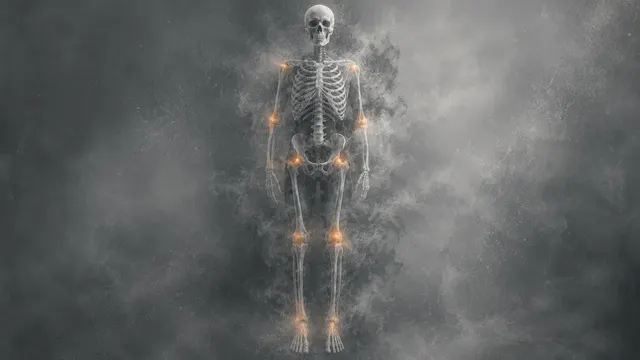
Whole-body bone scan read: metastatic hotspots, Bone Scan Index, PCWG3 progression and ATTR-CM flag.
Nuclear Cardiology (MPI and Cardiac Amyloid)
Nuclear cardiology read: perfusion scores, gated function, myocardial blood flow and ATTR-CM grade.
Lung V/Q Scintigraphy
Lung V/Q read: segmental mismatched perfusion defects and a pulmonary embolism call per EANM 2019.
Renal Scintigraphy
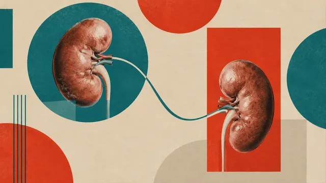
Renal scintigraphy read: diuretic renogram curves, split function, drainage and DMSA cortical scars.