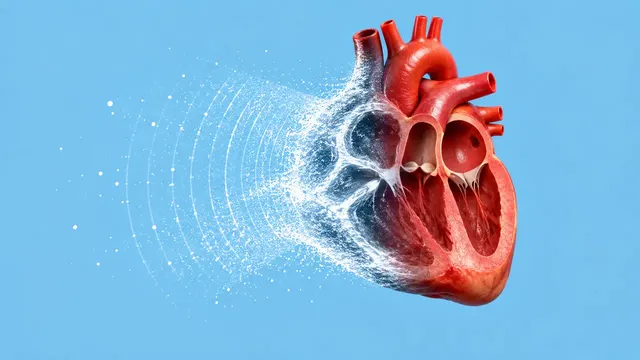
Echocardiography (TTE)
Full TTE read: views, LVEF, chambers, diastology, valves and effusion as ASE measurements.
Cardiac15 sections
US· cine
Organ-by-organ abdominal read: liver, gallbladder, kidneys, spleen, aorta and paediatric bowel.
This model is not available to run yet. Vote to show interest and get an email when it opens.
Abdominal and Renal Ultrasound reads complete and limited abdominal studies: right upper quadrant, renal, aortic and right-lower-quadrant scans, in adults and children. The run recognises which kind of study was uploaded and which standard plane each image shows, so a renal-colic scan and a full abdomen both come back organised by organ.
The result goes organ by organ. The liver gets a steatosis grade with the hepatorenal index and a focal-lesion list with a surveillance category; the gallbladder gets stones, sludge, wall, pericholecystic fluid, polyps and the common bile duct; the kidneys get hydronephrosis grade, length, cortex, cysts and stones; the spleen and aorta are measured. For children, the paediatric acute-abdomen questions are answered: appendicitis, intussusception and pyloric stenosis.
Decision support for the physician who performs or reads abdominal ultrasound: right-upper-quadrant pain, abnormal liver tests, renal colic or acute kidney injury, HCC surveillance, AAA screening and the child with abdominal pain. Organ measurements depend on the plane, and adult and paediatric norms differ, so each value names its image and reference.
Radiologists and radiology residents, internists, gastroenterologists and hepatologists, nephrologists and urologists, emergency physicians and paediatric radiologists. Developers add the run to reporting systems, liver surveillance programmes and AAA screening workflows.
| Requirement | Why |
|---|---|
SequenceOfUltrasoundRegions calibration |
Kidney length, CBD and aortic diameters, polyp and pylorus sizes in millimetres |
| Liver and right kidney in one image | The hepatorenal index compares their echogenicity |
| Transverse and longitudinal aorta | The maximum outer-to-outer diameter is taken across both |
| Spleen long-axis image | Spleen length |
| Compression cine of the right lower quadrant | Appendix compressibility cannot be judged on a still |
Bowel gas and body habitus limit views; organs not seen are listed with the reason rather than reported as normal.
Age (adult or paediatric norms), sex, the clinical question and risk context such as cirrhosis under HCC
surveillance go in clinical_context.
One JSON result in sections, ordered by organ, with findings, measurements and classifications. Organ masks
are returned as DICOM SEG and focal lesions keep a lesion_id and liver location.
| Section | Content |
|---|---|
| Organ segmentation | Standard-plane label per image; liver, gallbladder, kidney (capsule, cortex, medulla, central echo complex) and spleen masks |
| Liver steatosis | Steatosis grade (none, mild, moderate, severe) and hepatorenal index |
| Focal liver lesions | Lesion detection, cystic or solid, size and LI-RADS US surveillance category |
| Gallbladder and bile duct | Stones, sludge, wall thickness, pericholecystic fluid, distension, polyps with size, CBD diameter and a gallbladder-cancer flag |
| Hydronephrosis | Grade (none, mild, moderate, severe) and side |
| Kidneys | Length, cortical thickness and echogenicity, simple or complex cysts, stones |
| Spleen size | Length and a splenomegaly flag |
| Abdominal aorta | Maximum outer-to-outer diameter, AAA flag and mural thrombus |
| Appendicitis | Appendix seen or not, diameter, compressibility, periappendiceal fat or fluid, appendicolith |
| Intussusception | Target or pseudokidney sign, ileocolic or small-bowel type, length |
| Pyloric stenosis | Pyloric muscle thickness, channel length and gastric outlet passage |
| Draft report | Opt-in (options.report): an English narrative that interprets the result for the reader (findings, impression, limitations); every number is checked against the findings |
Gallbladder findings are grouped as the Tokyo 2018 local signs of acute cholecystitis: wall thickening, distension, stones or debris and pericholecystic fluid. The sonographic Murphy sign is a bedside finding that still images do not show. Polyps are placed in the SRU 2022 risk categories from their size and shape. An abdominal aortic diameter of 3.0 cm or more is flagged as an aneurysm, as defined in the SVS 2018 guideline.
us/fast reads trauma windows for free fluid.us/scrotal adds testicular torsion to the paediatric acute-abdomen work-up.One run returns every section its input supports.
Echocardiography (TTE)
Full TTE read: views, LVEF, chambers, diastology, valves and effusion as ASE measurements.
Obstetric Ultrasound
Trimester-aware pregnancy scan read: planes, biometry, GA, EFW, centiles, fluid and Doppler.
Thyroid and Neck Ultrasound
Thyroid nodule list with ACR TI-RADS level and FNA advice, gland volume and cervical lymph nodes.
Breast Ultrasound
Breast lesion list with BI-RADS v2025 descriptors and final assessment, axillary nodes and ABUS.
eFAST Trauma Ultrasound
eFAST read per window: free fluid, pericardial effusion, pneumothorax and an overall result.
Gynaecological Pelvic Ultrasound
Gynaecological pelvic read: O-RADS adnexal masses, endometrium, uterus and fibroids, follicles.