
Obstetric Ultrasound
Trimester-aware pregnancy scan read: planes, biometry, GA, EFW, centiles, fluid and Doppler.
Fetal14 sections
US· cine
Full TTE read: views, LVEF, chambers, diastology, valves and effusion as ASE measurements.
This model is not available to run yet. Vote to show interest and get an email when it opens.
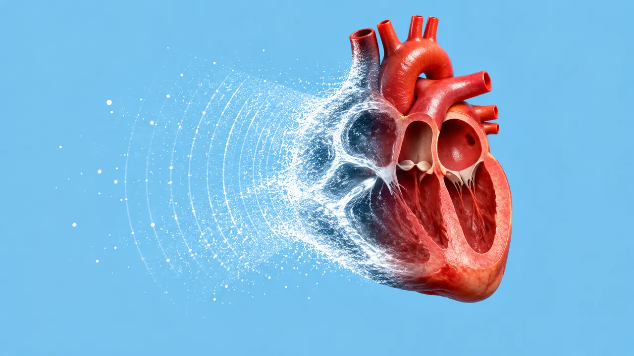
Echocardiography (TTE) reads a complete transthoracic study. Upload the 30 to 150 clips an echo cart exports, with 2D, M-mode, colour and PW, CW or tissue Doppler mixed together; the run labels every clip by view and mode, scores its quality and runs each measurement only on the clips that support it.
The result is an ASE-structured measurement table with findings per structure: ejection fraction and volumes, LV dimensions and mass, left atrial volume, right ventricular function, strain, regional wall motion, diastolic grade, valve severity, pulmonary hypertension probability, pericardial effusion and cardiomyopathy patterns. A focused section answers the four FoCUS questions from one to five handheld clips.
Decision support for the first read of an echo: the fellow or sonographer preparing a study for sign-off, the on-call physician who needs an ejection fraction and an effusion answer at night, and the emergency or ICU doctor scanning with a handheld probe. Each value carries its source clip and beat count.
Cardiologists, echo sonographers, cardiology fellows, emergency and intensive-care physicians and cardiac anaesthesiologists. Developers add the same run to a PACS, an echo reporting system or a heart-failure registry.
| Requirement | Why |
|---|---|
Cine with FrameTime or FrameTimeVector, CineRate and NumberOfFrames |
EF, strain and wall motion need frame timing; stills are refused for them |
SequenceOfUltrasoundRegions |
Calibrates millimetres per region and the velocity axis of each Doppler strip |
HeartRate and the ECG trace when present |
End-diastole and end-systole come from the ECG or from the LV area curve |
| Apical 4- and 2-chamber views (3-chamber for strain) | Biplane LV and LA volumes need both apical views; GLS uses all three |
| 3 to 5 beats on each Doppler strip | Envelopes are traced beat by beat and averaged; more beats in atrial fibrillation |
Handheld MP4 carries no calibration, so its millimetre values depend on a readable depth scale or a user-entered depth, and the result names the scale source.
Height and weight for BSA indexing, sex (ASE normal ranges are sex-specific), age, heart rhythm and the clinical
question go in clinical_context. Mark contrast (LVO) studies and prosthetic valves there as well.
One JSON result in sections, with findings, measurements, classifications and a clips[] list giving view,
mode, quality and cardiac phases for every clip. A measurement records whether it was computed, read from on-screen
calipers or taken from vendor SR. A foreshortened or off-axis clip yields "insufficient view" and its reason.
| Section | Content |
|---|---|
| Views and image quality | View (PLAX, RV inflow, PSAX levels, A4C/A5C/A2C/A3C, subcostal, SSN), mode and quality score per clip |
| Chamber segmentation | LV endocardium and epicardium, LA, RV and RA masks on every frame, with areas and volumes |
| LVEF and volumes | Biplane Simpson LVEF with a video-based estimate, EDV and ESV (indexed), beat-to-beat spread, HF category |
| LV size and mass | IVSd, LVIDd/s, LVPWd, LV mass and index, relative wall thickness and LV geometry |
| Left atrial volume | Biplane LA volume at end-systole, LAVi and LA diameter |
| Right ventricular function | TAPSE, fractional area change, S′, RV basal diameter and RV/LV ratio |
| Global longitudinal strain | GLS (%) and the segmental bull's-eye |
| Regional wall motion | Per-segment grade (normal, hypokinetic, akinetic, dyskinetic), WMSI and coronary territory |
| Diastolic function | E, A, E/A, septal and lateral e′, E/e′, TR Vmax, LAVi, diastolic grade and LA pressure estimate |
| Valve disease | AS (Vmax, mean gradient, AVA by continuity, DVI); MR, AR, TR (vena contracta, PISA EROA, jet); MS gradient; AS screen from 2D-only PLAX/PSAX |
| Pulmonary hypertension probability | TR Vmax, RVSP, IVC size and collapse (RAP), RA area, PA acceleration time and echo probability |
| Pericardial effusion | Presence, size (small, moderate, large) and tamponade signs (RA/RV collapse, plethoric IVC) |
| Cardiomyopathy patterns | HCM versus cardiac amyloidosis versus other LVH, LVOT obstruction, dilated pattern and next-test guidance |
| Focused cardiac ultrasound | LV function with EF estimate, RV dilatation (RV/LV > 1), effusion with tamponade signs, IVC size and collapse |
| Draft report | Opt-in (options.report): an English narrative that interprets the result for the reader (findings, impression, limitations); every number is checked against the findings |
The HF category follows LVEF: reduced at 40 % or less, mildly reduced at 41–49 %, preserved at 50 % or more. LAVi above 34 mL/m² is enlarged per ASE 2015. Each section records the guideline version its grade came from. The read is built for adult TTE; paediatric and congenital studies are read for ejection fraction only.
us/lung and us/vascular complete the breathless-patient work-up at the bedside.us/echo-tee reads transoesophageal studies.One run returns every section its input supports.
Obstetric Ultrasound
Trimester-aware pregnancy scan read: planes, biometry, GA, EFW, centiles, fluid and Doppler.
Thyroid and Neck Ultrasound
Thyroid nodule list with ACR TI-RADS level and FNA advice, gland volume and cervical lymph nodes.
Abdominal and Renal Ultrasound
Organ-by-organ abdominal read: liver, gallbladder, kidneys, spleen, aorta and paediatric bowel.
Breast Ultrasound
Breast lesion list with BI-RADS v2025 descriptors and final assessment, axillary nodes and ABUS.
eFAST Trauma Ultrasound
eFAST read per window: free fluid, pericardial effusion, pneumothorax and an overall result.
Gynaecological Pelvic Ultrasound
Gynaecological pelvic read: O-RADS adnexal masses, endometrium, uterus and fibroids, follicles.