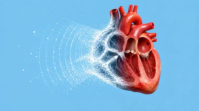
Echocardiography (TTE)
Full TTE read: views, LVEF, chambers, diastology, valves and effusion as ASE measurements.
Cardiac15 sections
US· cine
eFAST read per window: free fluid, pericardial effusion, pneumothorax and an overall result.
This model is not available to run yet. Vote to show interest and get an email when it opens.
eFAST Trauma Ultrasound reads the extended focused assessment with sonography for trauma performed in the primary survey: the right upper quadrant (hepatorenal space and right thorax), the left upper quadrant (splenorenal space and left thorax), the pelvis, the subxiphoid pericardial window and both anterior chest walls. Upload the four to six window clips, often exported as MP4 from a handheld device.
Each window comes back positive, negative or indeterminate, with the reason. Free fluid is located and sized, pericardial fluid is checked for tamponade signs, and the anterior chest clips are read for lung sliding and lung point. An overall eFAST result and next-step guidance sum up the scan.
Decision support for the emergency or trauma physician who has just scanned an injured patient, and a structured record of the scan for the trauma chart and for teaching. Novices miss small volumes, and fluid-filled bowel or perinephric fat can mimic blood, so each positive window shows its evidence frame.
Emergency physicians, trauma surgeons, emergency medicine residents, prehospital and military clinicians who record eFAST clips, and ultrasound educators reviewing trainee scans. Developers add the run to handheld-ultrasound apps and trauma registries.
| Requirement | Why |
|---|---|
| Cine clips | Lung sliding exists only over time, and a sweep through each window shows fluid that one frame can miss |
| Landmarks in view (liver tip, kidney pole, diaphragm, bladder, heart) | A window is adequate only when its required landmarks are seen; otherwise it is indeterminate |
| One clip per anterior hemithorax | Pneumothorax is reported for each side |
Obesity and subcutaneous emphysema degrade windows; the result names the window that failed and why. Size classes for fluid use the depth scale when one is readable.
The clinical question, for example blunt trauma in a hypotensive patient, goes in clinical_context and frames
the next-step guidance.
One JSON result in sections with a per-window verdict table, findings, quality and fluid masks on the evidence
frames, followed by the overall eFAST result. Indeterminate windows are listed with their reason.
| Section | Content |
|---|---|
| Windows and adequacy | Window per clip (RUQ, LUQ, pelvis longitudinal and transverse, subxiphoid, left and right chest) and whether its required landmarks are seen |
| Free fluid | Anechoic free fluid by location (hepatorenal, perisplenic, pelvic, pleural or haemothorax) with a size class |
| Pericardial effusion | Pericardial fluid on the subxiphoid or parasternal view, with tamponade flags |
| Pneumothorax | Lung sliding and lung point on each anterior chest wall, with a pneumothorax probability |
| Draft report | Opt-in (options.report): an English narrative that interprets the result for the reader (findings, impression, limitations); every number is checked against the findings |
Fluid in Morison's pouch, the splenorenal recess or the pelvis is reported with its location, because the location points to the injured organ. The pneumothorax read is the same one used in lung ultrasound, applied to the trauma windows.
us/lung reads a full lung ultrasound protocol.us/echo (focused section) and us/abdomen (aorta) extend an undifferentiated-shock work-up.One run returns every section its input supports.
Echocardiography (TTE)
Full TTE read: views, LVEF, chambers, diastology, valves and effusion as ASE measurements.
Obstetric Ultrasound
Trimester-aware pregnancy scan read: planes, biometry, GA, EFW, centiles, fluid and Doppler.
Thyroid and Neck Ultrasound
Thyroid nodule list with ACR TI-RADS level and FNA advice, gland volume and cervical lymph nodes.
Abdominal and Renal Ultrasound
Organ-by-organ abdominal read: liver, gallbladder, kidneys, spleen, aorta and paediatric bowel.
Breast Ultrasound
Breast lesion list with BI-RADS v2025 descriptors and final assessment, axillary nodes and ABUS.
Gynaecological Pelvic Ultrasound
Gynaecological pelvic read: O-RADS adnexal masses, endometrium, uterus and fibroids, follicles.