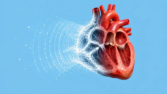
Echocardiography (TTE)
Full TTE read: views, LVEF, chambers, diastology, valves and effusion as ASE measurements.
Cardiac15 sections
US· cine
Thyroid nodule list with ACR TI-RADS level and FNA advice, gland volume and cervical lymph nodes.
This model is not available to run yet. Vote to show interest and get an email when it opens.
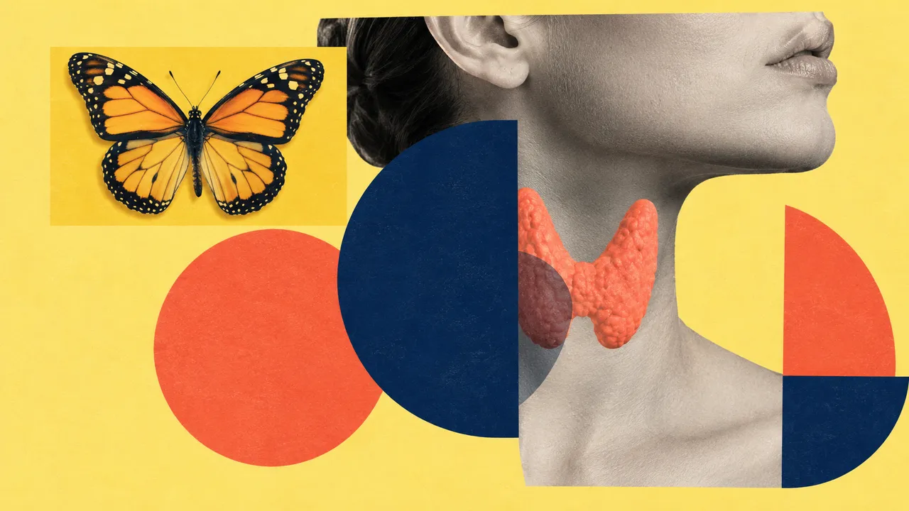
Thyroid and Neck Ultrasound reads a thyroid study: stills or short cine loops of both lobes and the isthmus, a transverse and a longitudinal view of each nodule, and images of the neck nodes. Lobe and plane labels typed on the screen ("LT LOBE TRANS", "SAG") are read as metadata, so the run knows which image shows which nodule and in which plane.
It builds one nodule list for the study. Each nodule is detected and outlined, matched across its transverse and longitudinal images so it counts once, measured in three axes and scored feature by feature with ACR TI-RADS 2017. The points give a TR level, and the nodule's size against that level gives the recommendation: fine-needle aspiration, follow-up or no further action. Lobe volumes and suspicious neck nodes complete the read.
Decision support for the reader of a thyroid ultrasound, where risk scoring is a known source of variation between readers and of aspirations that were not needed. The result shows every feature and point behind a TR level, so the reader can check the descriptor that decided it.
Radiologists, endocrinologists, ENT and endocrine surgeons, and sonographers who prepare thyroid worksheets. Developers call the run to fill TI-RADS fields in a reporting system or to triage nodule clinics.
| Requirement | Why |
|---|---|
| Transverse and longitudinal view of each nodule | Shape (taller-than-wide) and three-axis size need both planes |
SequenceOfUltrasoundRegions calibration |
TI-RADS recommendations depend on size in centimetres; uncalibrated images give features without size advice |
| Unobstructed nodule margins | Burned-in calipers drawn over a margin can hide it; such margins are flagged |
| Lateral and central neck images | Needed for the lymph-node section |
Age, sex and the clinical question (incidental nodule, known goitre, follow-up of a previously scored nodule) go in
clinical_context.
One JSON result in sections with findings, measurements and classifications. Each nodule keeps a stable
lesion_id and a location (lobe, upper, mid or lower third, isthmus), and its outline is returned as DICOM SEG.
| Section | Content |
|---|---|
| Nodules | Detection, outline and three-axis size of every nodule, with location and the images it appears on |
| ACR TI-RADS | Composition, echogenicity, shape, margin and echogenic foci; points, TR1–TR5 level and the FNA or follow-up recommendation by size |
| Thyroid volume | Lobe and isthmus segmentation, ellipsoid volumes per lobe and a goitre flag |
| Cervical lymph nodes | Node detection with suspicious features: microcalcifications, cystic change, hyperechoic cortex, peripheral vascularity, round shape |
| Draft report | Opt-in (options.report): an English narrative that interprets the result for the reader (findings, impression, limitations); every number is checked against the findings |
Points from the five feature categories are summed: 0 points is TR1, 2 is TR2, 3 is TR3, 4 to 6 is TR4 and 7 or more is TR5. Large comet-tail artefacts score 0 and punctate echogenic foci score 3, so the run separates them explicitly; this is a frequent point of disagreement between readers.
| Level | FNA if the nodule is | Follow-up if the nodule is |
|---|---|---|
| TR3 | 2.5 cm or more | 1.5 cm or more |
| TR4 | 1.5 cm or more | 1.0 cm or more |
| TR5 | 1.0 cm or more | 0.5 cm or more |
A TR level without calibrated size carries features and points but no size-based recommendation.
One run returns every section its input supports.
Echocardiography (TTE)
Full TTE read: views, LVEF, chambers, diastology, valves and effusion as ASE measurements.
Obstetric Ultrasound
Trimester-aware pregnancy scan read: planes, biometry, GA, EFW, centiles, fluid and Doppler.
Abdominal and Renal Ultrasound
Organ-by-organ abdominal read: liver, gallbladder, kidneys, spleen, aorta and paediatric bowel.
Breast Ultrasound
Breast lesion list with BI-RADS v2025 descriptors and final assessment, axillary nodes and ABUS.
eFAST Trauma Ultrasound
eFAST read per window: free fluid, pericardial effusion, pneumothorax and an overall result.
Gynaecological Pelvic Ultrasound
Gynaecological pelvic read: O-RADS adnexal masses, endometrium, uterus and fibroids, follicles.