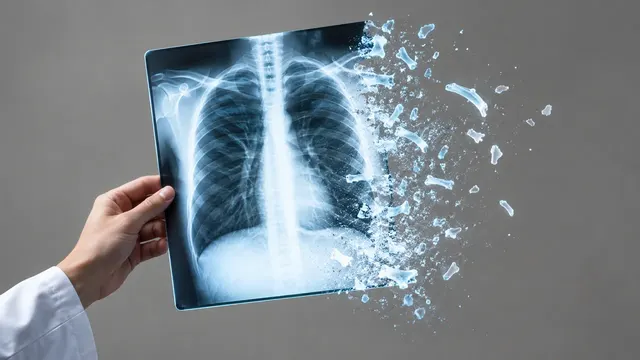
Chest radiograph read: urgent flags, findings with boxes, device tips, CTR and interval change.
What it does
Chest X-ray reads a chest radiograph study and puts urgent findings first. Upload a frontal film (PA or AP), with a lateral view and a prior study when you have them. The run checks projection and exposure, flags pneumothorax, malpositioned tubes, large effusions, consolidation and subdiaphragmatic free air, then returns the full finding list.
Local findings come with boxes on the image; pneumothorax, effusion and consolidation come with size or extent; every endotracheal tube, nasogastric tube, central venous catheter and Swan-Ganz catheter comes with its tip position and, for the endotracheal tube, the tip-to-carina distance in millimetres. The cardiothoracic ratio, a comparison with the prior film and two opportunistic risk estimates complete the read.
Intended use
Decision support for the doctor who reads the film, often without a radiologist: an ED physician at night, an ICU team checking lines on the morning round, a GP, or a TB screening programme. Each finding comes with its evidence, and the guidance points to the matching pathway (BTS pneumothorax management, CT for a nodule, molecular testing for a TB-positive screen).
Who it is for
Emergency, intensive-care and respiratory physicians, radiologists and residents, GPs and TB programme staff. Children are handled inside this model through age-aware routing; neonatal films belong to NICU Radiograph. Developers call the same run to add chest X-ray analysis to a PACS, worklist or screening app.
Accepted input
- DICOM radiographs: CR and DX (For Presentation or For Processing) objects; one frontal image is enough.
- Studies: PA or AP frontal, optional lateral, and an optional prior study for the comparison section.
- PNG or JPEG for development: accepted, but marked uncalibrated, so no millimetre values are returned.
Requirements
| Requirement | Why |
|---|---|
| A frontal view (PA or AP) | Every section starts from the frontal film; the lateral and prior add to it |
ImagerPixelSpacing or PixelSpacing |
Needed for tube-to-carina distance, nodule diameter and pneumothorax rim distance in mm |
ViewPosition and patient position |
AP supine films overestimate heart size and hide anterior pneumothorax; the ratio is flagged as unreliable |
PatientAge or birth date |
Selects the paediatric path below 18 years and the TB screening age (15 years and over) |
MONOCHROME1 images are inverted automatically; rotation, poor inspiration, clipping and skin folds are reported in the quality section.
Optional context
Age, sex and smoking history feed the risk sections. The indication or clinical question (for example "ARDS?" or
"line check") in clinical_context focuses the guidance.
The result
One JSON result in sections, with device positions, calibration and projection quality as structured fields.
| Section | Content |
|---|---|
| Triage | Normal or abnormal, plus urgent flags for worklist priority |
| Image quality | Projection (PA, AP, lateral), rotation, inspiration, penetration, clipping, collimation and artefacts |
| Findings | Multi-label probabilities for common chest findings, with boxes for local findings |
| Pneumothorax | Presence, side, mask, rim distance, tension signs and chest-drain presence |
| Lines and tubes | ETT, NGT, CVC and Swan-Ganz tips, carina landmark, ETT–carina distance and a normal, borderline or abnormal position |
| Pneumonia | Airspace opacity and consolidation masks with probability, lobe and laterality |
| Tuberculosis | TB abnormality score 0–100 against a locally calibrated threshold, with boxes on TB-consistent findings |
| Lung nodules | Nodule and mass boxes, diameter in mm, calcified or not, and a CT recommendation |
| Cardiothoracic ratio | Heart and thoracic widths from masks and the ratio on PA films |
| Pleural effusion | Side and size grade: costophrenic blunting, meniscus, or half the hemithorax or more |
| Pulmonary oedema | Severity 0–3 (none, vascular congestion, interstitial, alveolar); RALE score when ARDS is queried |
| Fractures | Rib, clavicle, scapula and vertebral-body fractures, acute or healed, with rib numbering |
| Anatomy segmentation | Masks of lungs, heart, clavicles, scapulae, mediastinum, aorta, diaphragm and spine |
| Comparison with prior | Each finding improving, stable or worsening; new and resolved findings; device moves |
| Cardiovascular risk | 10-year cardiovascular risk and a radiographic biological age |
| Lung cancer risk | Long-term lung-cancer risk and a low-dose CT screening suggestion |
| Draft report | Opt-in (options.report): an English narrative that interprets the result for the reader (findings, impression, limitations); every number is checked against the findings |
Standards
- DICOM: Chest CAD SR (TID 4100) for findings, TID 1500 Measurement Report for CTR and ETT–carina distance, TID 4019 Algorithm Identification, SEG for lungs, heart, pneumothorax and devices, GSPS overlays, KOS key images and an Encapsulated PDF; display through the IHE AI Results (AIR) profile.
- Terminology: Fleischner Society glossary (a nodule is 3 cm or less, a mass is larger), RadLex and SNOMED CT.
- Guidelines: BTS pleural disease guideline (2023); IDSA/ATS 2019 community-acquired pneumonia guideline; WHO consolidated TB guidelines, Module 2, and the WHO TB CAD target product profile; Berlin ARDS definition.
- Risk comparators: ACC/AHA pooled cohort equations; USPSTF 2021 low-dose CT eligibility, with Lung-RADS applying on the follow-up CT.
Measurement notes
Pneumothorax size is classed by two conventions: a large pneumothorax has a rim of 2 cm or more at the level of the hilum (BTS) or 3 cm or more from apex to cupola (ACCP). The adult ETT tip target is about 5 ± 2 cm above the carina. A CTR above 0.5 on a PA film sets the cardiomegaly flag; AP films report the ratio but mark it unreliable.
Limitations and failure modes
- Sections in version 1.0.0: triage, findings, pneumothorax, pneumonia, lines and tubes, projection. Boxes for local findings, pneumothorax size and chest drains, tuberculosis, nodule size, the cardiothoracic ratio, effusion grade, oedema score, fractures with boxes and the comparison with a prior are listed as not assessable until they join the run.
- Adults only: the networks were trained on adult films; films of patients under 18 years are rejected.
- Small or anterior pneumothorax on supine AP films is found less often than an apical rim on an erect film, and the outline of a small pneumothorax can be partial; skin folds can mimic a pneumothorax edge.
- AP portable films enlarge the heart and mediastinum: cardiomegaly and a widened mediastinum read more often.
- Operating points were set on VinDr-CXR (one hospital network); on low-resolution or heavily processed exports the panel finds fewer subtle findings.
- Central line and nasogastric tips are given as a position category, not as a tip point; the endotracheal tube tip and the carina are points.
- One frontal film is read per study; the lateral film is not analysed yet.
Result sections
One run returns every section its input supports, at one price. Sections that cannot run say why (for example, not in input).
| Section | What it returns | Standards | Status |
|---|---|---|---|
Triage #triage | Worklist priority: critical for pneumothorax, a malpositioned endotracheal or nasogastric tube, free air or pneumomediastinum; urgent for effusion, consolidation, a widened mediastinum, a malpositioned central line or a nasogastric tip off the film; routine otherwise. Every flag points to its finding.findings | IHE AI Results (AIR) (Rev 1.3) | Since v1.0.0 |
Image quality #quality | Projection (PA, AP or lateral) of the read film, rotation of the stored image, calibration and exposure flags; rotation, inspiration and penetration of the patient join later.classifications | — | Since v1.0.0 |
Findings #findings | Normal or abnormal, and a 37-finding panel in Fleischner glossary terms: the 17 findings with a radiologist reference are always stated (present, absent, or for consolidation, nodule or mass and cardiomegaly also not excluded); the others are listed when present. Boxes for local findings join with the VinDr-trained detector.findings · artifacts | Fleischner Society glossary of terms for thoracic imaging (2008); DICOM SR TID 1500 (2026d) | Since v1.0.0 |
Pneumothorax #pneumothorax | Pneumothorax present or absent, with side and outline on the film. Rim size by the BTS and ACCP conventions and chest-drain detection join later.findings · guidance · artifacts | BTS Guideline for Pleural Disease (2023); DICOM Segmentation IOD (2026d) | Since v1.0.0 |
Lines and tubes #lines-tubes | Endotracheal tube, nasogastric tube, central venous catheter and Swan-Ganz catheter: presence, outline and the RANZCR position category (normal, borderline, abnormal; nasogastric tip not on the film); the endotracheal tube tip, the carina and the tip-to-carina distance in millimetres when the film carries a pixel spacing.findings · measurements · classifications · guidance · artifacts | RANZCR CLiP catheter and line position categories (2021); Endotracheal tube position (adult target 5 ± 2 cm above the carina) (1976) | Since v1.0.0 |
Pneumonia #pneumonia | Airspace opacity consistent with pneumonia, with side and outline; lobe joins later.findings · artifacts | IDSA/ATS community-acquired pneumonia guideline (2019) | Since v1.0.0 |
Draft report #report | Opt-in draft report (findings, impression, limitations) written from the structured result; every number is validated against it.report | — | Since v1.0.0 |
Tuberculosis #tuberculosis | TB abnormality score 0–100 with a locally set threshold for screening programmes (WHO CAD), after a bacteriologically confirmed evaluation.scores · findings | WHO consolidated guidelines on tuberculosis (2021) | Planned · P1 |
Lung nodules #lung-nodules | Nodule and mass boxes with diameter and a CT recommendation.findings · measurements · guidance | — | Planned · P1 |
Cardiothoracic ratio #cardiomegaly | Heart and thoracic widths and the cardiothoracic ratio on PA films.measurements | — | Planned · P1 |
Pleural effusion #effusion | Effusion side and size grade (blunting, meniscus, half the hemithorax or more).findings | BTS Guideline for Pleural Disease (2023) | Planned · P2 |
Pulmonary oedema #edema | Oedema severity 0–3 and the RALE score.scores | — | Planned · P2 |
Fractures #fractures | Rib, clavicle, scapula and vertebral fractures with boxes and rib numbers.findings | — | Planned · P2 |
Anatomy segmentation #segment-anatomy | Lungs, heart, clavicles, scapulae, mediastinum, aorta, diaphragm and spine outlines (lungs and heart already come with every read as evidence).artifacts | — | Planned · P2 |
Comparison with prior #compare | Per-finding change against a prior film (improving, stable, worsening), new and resolved findings, device moves. Requires: A prior chest film in prior_files.changes | — | Planned · P1 |
Cardiovascular risk #cvd-risk | Ten-year cardiovascular risk from the frontal film.risks | — | Planned · P3 |
Lung cancer risk #lung-cancer-risk | Long-term lung-cancer risk and the screening suggestion.risks | — | Planned · P3 |
Validated performance
Numbers come only from MedRun's own evaluation harness (make bench (models/xr-chest)), with dataset, split and date.
| Section | Metric | Value | Dataset | Date |
|---|---|---|---|---|
| findings | Abnormal vs normal AUC | 0.92495% CI 0.913–0.934 | VinDr-CXR (official test (5-radiologist consensus)) · n=2999 | 2026-10-14 |
| findings | Pleural effusion AUC | 0.99395% CI 0.99–0.996 | VinDr-CXR (official test) · n=2999 | 2026-10-14 |
| findings | Consolidation AUC | 0.97195% CI 0.963–0.979 | VinDr-CXR (official test) · n=2999 | 2026-10-14 |
| findings | Cardiomegaly AUC | 0.96495% CI 0.957–0.971 | VinDr-CXR (official test) · n=2999 | 2026-10-14 |
| findings | Nodule or mass AUC | 0.94295% CI 0.925–0.957 | VinDr-CXR (official test) · n=2999 | 2026-10-14 |
| findings | Atelectasis AUC | 0.96295% CI 0.953–0.972 | VinDr-CXR (official test) · n=2999 | 2026-10-14 |
| pneumonia | Pneumonia AUC | 0.96395% CI 0.954–0.971 | VinDr-CXR (official test) · n=2999 | 2026-10-14 |
| findings | Pleural effusion AUC | 0.91395% CI 0.873–0.951 | CheXpert (validation (frontal) · n=201 | 2026-10-14 |
| pneumothorax | Pneumothorax AUC | 0.95195% CI 0.913–0.979 | NIH ChestX-ray14 radiologist boxes (films not in SIIM-ACR (held out from both networks)) · n=688 | 2026-10-14 |
| pneumothorax | Pneumothorax AUC | 0.99395% CI 0.984–0.999 | VinDr-CXR (official test) · n=2999 | 2026-10-14 |
Pricing
| Endpoint | Unit | Price |
|---|---|---|
| xr/chest/interpret | request | $0.05 |
| Draft report (opt-in, options.report) | per run | +$0.02 |
- One price per study, whatever the number of films.
- Rejected inputs (for example, no chest film in the upload, or a child's film) and failed runs are not charged.
Changelog
- v1.0.0Oct 14, 2026major
- First MedRun build of the chest radiograph read; triage, findings panel, pneumothorax, pneumonia, lines and tubes with the ETT–carina distance, DICOM SEG and SR.