Musculoskeletal X-ray
Bone and joint radiograph read: fractures, dislocations, bone age, osteoarthritis and alignment.
MSKPelvis11 sections
XR· image-2d
Chest radiograph read: urgent flags, findings with boxes, device tips, CTR and interval change.
This model is not available to run yet. Vote to show interest and get an email when it opens.
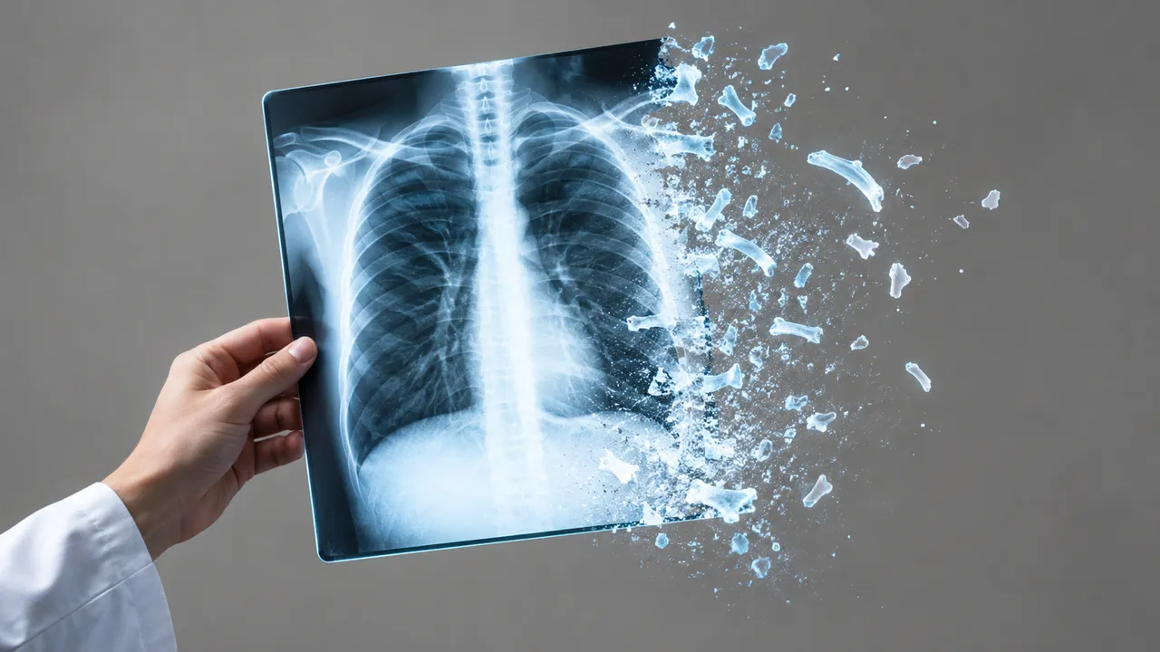
Chest X-ray reads a chest radiograph study and puts urgent findings first. Upload a frontal film (PA or AP), with a lateral view and a prior study when you have them. The run checks projection and exposure, flags pneumothorax, malpositioned tubes, large effusions, consolidation and subdiaphragmatic free air, then returns the full finding list.
Local findings come with boxes on the image; pneumothorax, effusion and consolidation come with size or extent; every endotracheal tube, nasogastric tube, central venous catheter and Swan-Ganz catheter comes with its tip position and, for the endotracheal tube, the tip-to-carina distance in millimetres. The cardiothoracic ratio, a comparison with the prior film and two opportunistic risk estimates complete the read.
Decision support for the doctor who reads the film, often without a radiologist: an ED physician at night, an ICU team checking lines on the morning round, a GP, or a TB screening programme. Each finding comes with its evidence, and the guidance points to the matching pathway (BTS pneumothorax management, CT for a nodule, molecular testing for a TB-positive screen).
Emergency, intensive-care and respiratory physicians, radiologists and residents, GPs and TB programme staff. Children are handled inside this model through age-aware routing; neonatal films belong to NICU Radiograph. Developers call the same run to add chest X-ray analysis to a PACS, worklist or screening app.
| Requirement | Why |
|---|---|
| A frontal view (PA or AP) | Every section starts from the frontal film; the lateral and prior add to it |
ImagerPixelSpacing or PixelSpacing |
Needed for tube-to-carina distance, nodule diameter and pneumothorax rim distance in mm |
ViewPosition and patient position |
AP supine films overestimate heart size and hide anterior pneumothorax; the ratio is flagged as unreliable |
PatientAge or birth date |
Selects the paediatric path below 18 years and the TB screening age (15 years and over) |
MONOCHROME1 images are inverted automatically; rotation, poor inspiration, clipping and skin folds are reported in the quality section.
Age, sex and smoking history feed the risk sections. The indication or clinical question (for example "ARDS?" or
"line check") in clinical_context focuses the guidance.
One JSON result in sections, with device positions, calibration and projection quality as structured fields.
| Section | Content |
|---|---|
| Triage | Normal or abnormal, plus urgent flags for worklist priority |
| Image quality | Projection (PA, AP, lateral), rotation, inspiration, penetration, clipping, collimation and artefacts |
| Findings | Multi-label probabilities for common chest findings, with boxes for local findings |
| Pneumothorax | Presence, side, mask, rim distance, tension signs and chest-drain presence |
| Lines and tubes | ETT, NGT, CVC and Swan-Ganz tips, carina landmark, ETT–carina distance and a normal, borderline or abnormal position |
| Pneumonia | Airspace opacity and consolidation masks with probability, lobe and laterality |
| Tuberculosis | TB abnormality score 0–100 against a locally calibrated threshold, with boxes on TB-consistent findings |
| Lung nodules | Nodule and mass boxes, diameter in mm, calcified or not, and a CT recommendation |
| Cardiothoracic ratio | Heart and thoracic widths from masks and the ratio on PA films |
| Pleural effusion | Side and size grade: costophrenic blunting, meniscus, or half the hemithorax or more |
| Pulmonary oedema | Severity 0–3 (none, vascular congestion, interstitial, alveolar); RALE score when ARDS is queried |
| Fractures | Rib, clavicle, scapula and vertebral-body fractures, acute or healed, with rib numbering |
| Anatomy segmentation | Masks of lungs, heart, clavicles, scapulae, mediastinum, aorta, diaphragm and spine |
| Comparison with prior | Each finding improving, stable or worsening; new and resolved findings; device moves |
| Cardiovascular risk | 10-year cardiovascular risk and a radiographic biological age |
| Lung cancer risk | Long-term lung-cancer risk and a low-dose CT screening suggestion |
| Draft report | Opt-in (options.report): an English narrative that interprets the result for the reader (findings, impression, limitations); every number is checked against the findings |
Pneumothorax size is classed by two conventions: a large pneumothorax has a rim of 2 cm or more at the level of the hilum (BTS) or 3 cm or more from apex to cupola (ACCP). The adult ETT tip target is about 5 ± 2 cm above the carina. A CTR above 0.5 on a PA film sets the cardiomegaly flag; AP films report the ratio but mark it unreliable.
One run returns every section its input supports.
Musculoskeletal X-ray
Bone and joint radiograph read: fractures, dislocations, bone age, osteoarthritis and alignment.
Abdominal X-ray
Abdominal film read: bowel gas pattern, free air, radio-opaque stones and swallowed objects.
NICU Radiograph
Neonatal babygram read: umbilical line and tube tips by vertebral level, plus NEC signs.
Spine X-ray
Spine radiograph read: scoliosis curves, vertebral fractures, sagittal balance and slip grades.
DXA (Bone Densitometry)
DXA read: T-scores by ISCD rules, VFA fractures, aortic calcification and body composition.
Hysterosalpingogram
HSG read: fill and spill for each fallopian tube, with occlusion level and hydrosalpinx.