Chest X-ray
Chest radiograph read: urgent flags, findings with boxes, device tips, CTR and interval change.
Chest17 sections
XR· image-2d
Chest, bone, spine and abdominal radiographs and fluoroscopy.
9 models
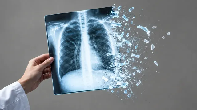
Chest X-ray
Chest radiograph read: urgent flags, findings with boxes, device tips, CTR and interval change.
Musculoskeletal X-ray
Bone and joint radiograph read: fractures, dislocations, bone age, osteoarthritis and alignment.
Abdominal X-ray
Abdominal film read: bowel gas pattern, free air, radio-opaque stones and swallowed objects.
NICU Radiograph
Neonatal babygram read: umbilical line and tube tips by vertebral level, plus NEC signs.
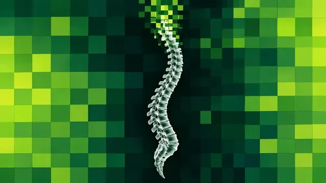
Spine X-ray
Spine radiograph read: scoliosis curves, vertebral fractures, sagittal balance and slip grades.
DXA (Bone Densitometry)
DXA read: T-scores by ISCD rules, VFA fractures, aortic calcification and body composition.
Hysterosalpingogram
HSG read: fill and spill for each fallopian tube, with occlusion level and hydrosalpinx.
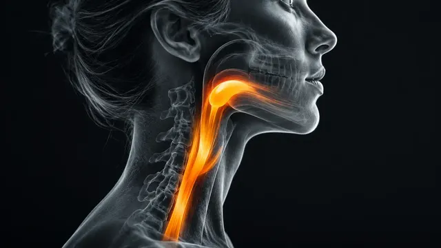
Swallow Study (VFSS)
Videofluoroscopic swallow read: PAS per bolus, aspiration frames, hyoid and UES motion, residue.
Voiding Cystourethrogram
Voiding cystourethrogram read: reflux side and International Reflux Study grade I–V.