Prostate Biopsy Pathology
Prostate core biopsy read: cancer per core, Gleason and ISUP grade, % pattern 4, tumour length.
Pelvis4 sections
WSI· wsi
Peripheral blood smear read: classified white cells, differential percentages and a blast alert.
This model is not available to run yet. Vote to show interest and get an email when it opens.
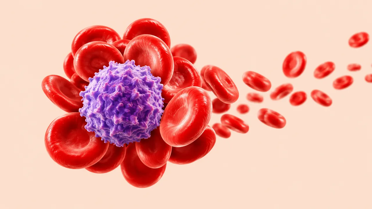
Peripheral Blood Smear reads the nucleated cells of a stained blood film. Smear review follows an abnormal complete blood count or an analyser flag, and its most important job is safety: not missing blasts or abnormal lymphocytes among otherwise ordinary cells.
The run locates and classifies each nucleated cell as neutrophil, lymphocyte, monocyte, eosinophil, basophil, immature granulocyte, blast, erythroblast or atypical cell, and turns the counts into a differential in percent. Every cell appears in a gallery grouped by class, so the reviewer sees the cells behind each number. When blasts or abnormal lymphocytes are found, the result raises an alert with "refer for haematology review" guidance.
Decision support for the laboratory morphology review: a pre-classified differential with every cell in view, and a safety net that puts suspicious smears first. Red-cell and platelet morphology are not graded by this model.
Haematology laboratory scientists and technologists, haematologists, clinical pathologists, and labs without a digital morphology analyser. Developers use the run to add a structured differential and an abnormal-cell alert to a laboratory information system.
Two kinds of input describe the same smear:
| Requirement | Why |
|---|---|
| Romanowsky-type stain (Wright, MGG) | Granules, nuclear chromatin and cytoplasm colour separate the cell classes |
| Readable monolayer on whole slides | Cells are classified where they lie singly, not in thick or feathered areas |
| Pixel spacing on whole slides | Cell size helps separate blasts from lymphocytes and monocytes |
The full blood count, analyser flags, age and the clinical question can be passed in clinical_context; the
guidance reads the differential against them.
| Section | Content |
|---|---|
| WBC differential | Class of every nucleated cell, differential percentages, cell gallery by class and the blast or abnormal-cell alert |
| Draft report | Opt-in (options.report): an English narrative that interprets the result for the reader (findings, impression, limitations); every number is checked against the findings |
Counts and percentages sit in measurements; each gallery cell keeps a link to its position on the slide or to its
source image.
DiagnosticReport with one LOINC-coded Observation per cell class.The alert names the cells classed as blasts or abnormal lymphocytes, shows them in their own gallery and carries the "refer for haematology review" guidance.
When blasts are flagged, Bone Marrow Aspirate (path/bone-marrow) reads the follow-up aspirate with a full
myelogram and blast percentage.
One run returns every section its input supports.
Prostate Biopsy Pathology
Prostate core biopsy read: cancer per core, Gleason and ISUP grade, % pattern 4, tumour length.
Any-Organ H&E Slide Analysis
Organ-agnostic H&E read: slide QC, tumour map, nuclei and mitoses, primary-site ranking, embeddings.
Breast Pathology
Breast case read from H&E, IHC and node slides: type, grade, biomarker scores, TILs and pN.
Colorectal Pathology
Colorectal polyp and cancer read: adenoma or serrated histology, dysplasia, MSI, tissue map.
Lung Cancer Pathology
Lung biopsy and resection read: NSCLC subtype, IASLC grade, PD-L1 TPS and EGFR mutation probability.
Bone Marrow Aspirate
Marrow aspirate read: cell-by-cell myelogram, blast percentage, M:E ratio and dysplasia flags.