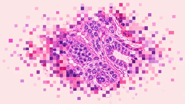
Any-Organ H&E Slide Analysis
Organ-agnostic H&E read: slide QC, tumour map, nuclei and mitoses, primary-site ranking, embeddings.
Whole body8 sections
WSI· wsi
Prostate core biopsy read: cancer per core, Gleason and ISUP grade, % pattern 4, tumour length.
This model is not available to run yet. Vote to show interest and get an email when it opens.
Prostate Biopsy Pathology reads a whole needle-biopsy case. A systematic biopsy often yields 10 to 12 or more cores, each scanned as an H&E whole-slide image; the run takes every core of the case together, so the result answers the questions of the sign-out: which cores carry carcinoma, how much, and of what grade.
For each core the result gives a carcinoma probability with a heatmap, the tumour length in millimetres and the percentage of the core involved. Cancer-bearing cores receive a Gleason pattern map, Gleason score, ISUP grade group 1–5, the percentage of pattern 4 and 5, and a cribriform flag. Core results then roll up into a case-level summary ordered by part. Prostatectomy slides are accepted too. A biochemical-recurrence risk score with a time-to-event estimate completes the result.
Decision support and a second read before sign-out: catching a small focus in a long core, checking a Gleason 3+4 against 4+3, or quantifying pattern 4 for an active-surveillance decision. Benign mimickers (atrophy, adenosis, high-grade PIN, seminal vesicle), treated prostate after radiation or hormonal therapy, and crushed or thin cores are the known pitfalls of the read, and quality flags mark crushed or thin cores.
General surgical pathologists without uropathology fellowship training, pathology residents, and small labs that lack a second reader. Developers call the same run to add prostate grading to a slide-management system or LIS.
| Requirement | Why |
|---|---|
| Scan at 20× or 40× | Gland architecture for Gleason patterns is read at this magnification |
| Pixel spacing in µm/px (about 0.5 at 20×, 0.25 at 40×) | Tumour length in mm comes from calibrated spacing |
| Stain recorded in the slide metadata or the upload | Routes the slide to the H&E read |
| Focused, unfolded tissue | Blur, folds and pen marks lower confidence and are listed in quality flags |
The indication (first biopsy, active surveillance, prior treatment) and clinical history can go in
clinical_context; they shape the guidance and the draft report.
One JSON result per case, organised in sections, with per-core and case-level values side by side.
| Section | Content |
|---|---|
| Cancer detection | Per core: carcinoma probability, heatmap, tumour length in mm and % of core involved |
| Gleason grading | Gleason pattern map, Gleason score, ISUP grade group 1–5, % pattern 4/5 and cribriform presence per core and per case |
| Recurrence risk | Biochemical-recurrence risk score and time-to-event estimate |
| Draft report | Opt-in (options.report): an English narrative that interprets the result for the reader (findings, impression, limitations); every number is checked against the findings |
DiagnosticReport with one Observation per score, and IHE PaLM APSR for LIS integration.The grade group follows from the Gleason score: 3+3 is group 1, 3+4 group 2, 4+3 group 3, any score of 8 group 4 and 9–10 group 5. Cores with grade group 2 or 3 also carry the percentage of pattern 4.
One run returns every section its input supports.
Any-Organ H&E Slide Analysis
Organ-agnostic H&E read: slide QC, tumour map, nuclei and mitoses, primary-site ranking, embeddings.
Breast Pathology
Breast case read from H&E, IHC and node slides: type, grade, biomarker scores, TILs and pN.
Colorectal Pathology
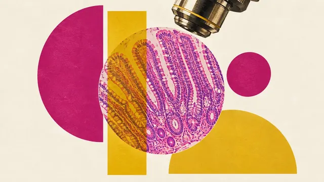
Colorectal polyp and cancer read: adenoma or serrated histology, dysplasia, MSI, tissue map.
Lung Cancer Pathology
Lung biopsy and resection read: NSCLC subtype, IASLC grade, PD-L1 TPS and EGFR mutation probability.
Peripheral Blood Smear
Peripheral blood smear read: classified white cells, differential percentages and a blast alert.
Bone Marrow Aspirate
Marrow aspirate read: cell-by-cell myelogram, blast percentage, M:E ratio and dysplasia flags.