Prostate Biopsy Pathology
Prostate core biopsy read: cancer per core, Gleason and ISUP grade, % pattern 4, tumour length.
Pelvis4 sections
WSI· wsi
Breast case read from H&E, IHC and node slides: type, grade, biomarker scores, TILs and pN.
This model is not available to run yet. Vote to show interest and get an email when it opens.
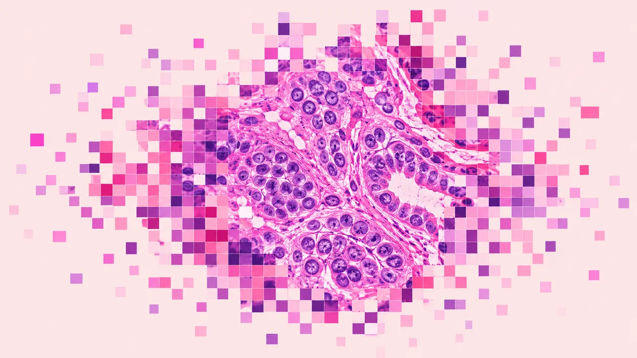
Breast Pathology reads a breast case the way it arrives at sign-out: the H&E slides of a core biopsy or excision, the ER, PR, HER2 and Ki-67 immunostains, and the sentinel or axillary lymph-node slides. Each slide is routed by its stain and marker, so one upload gives one case-level result.
On H&E the run classifies the lesion (benign, usual ductal hyperplasia, flat epithelial atypia, atypical ductal hyperplasia, DCIS, invasive carcinoma of no special type or invasive lobular carcinoma), scores the three Nottingham components and measures stromal TILs. On IHC it returns the HER2 score with membrane-staining percentages, ER and PR positivity with Allred and H-score, and the Ki-67 index. Node slides are screened for metastases, and the largest deposit sets the isolated-tumour-cell, micrometastasis or macrometastasis category.
Decision support for the biomarker panel and the node screen, where agreement between readers is weakest: HER2 null versus ultralow versus 1+, ER-low results, and small deposits in a long series of node slides. Each score is reported with its antibody clone, and the cell overlay shows what was counted.
Breast and general surgical pathologists, residents, and labs that score biomarkers manually. Developers use the run to add structured biomarker and nodal results to a LIS or a tumour-board workflow.
| Requirement | Why |
|---|---|
| Stain and marker per slide (and antibody clone for IHC) | Routes each slide to the right scoring and is reported with the score |
| Pixel spacing in µm/px | Deposit size in mm and mitotic counts per area need calibration |
| Part labels for node slides | Nodal results are reported per node |
Clinical history and prior treatment can be passed in clinical_context. Specimens after neoadjuvant
therapy are not graded for residual cancer burden.
One JSON result per case, with per-slide values and a case summary.
| Section | Content |
|---|---|
| Histologic type | Lesion category from benign through atypia and DCIS to invasive NST or lobular carcinoma |
| Nottingham grade | Tubule formation, nuclear pleomorphism and mitotic count, combined into grade 1–3 |
| ER and PR | % positive tumour nuclei, intensity distribution, Allred score, H-score and ER-low flag |
| HER2 score | 0 (null or ultralow), 1+, 2+ or 3+ with % complete and incomplete membrane staining and a HER2-low flag |
| Ki-67 index | Global and hot-spot index with a positive and negative cell overlay |
| Stromal TILs | Stromal TILs percentage with the tumour and stroma map it was measured on |
| Lymph node metastases | Per node: metastasis detected, largest deposit size and ITC, micrometastasis or macrometastasis |
| Draft report | Opt-in (options.report): an English narrative that interprets the result for the reader (findings, impression, limitations); every number is checked against the findings |
Observation per biomarker.One run returns every section its input supports.
Prostate Biopsy Pathology
Prostate core biopsy read: cancer per core, Gleason and ISUP grade, % pattern 4, tumour length.
Any-Organ H&E Slide Analysis
Organ-agnostic H&E read: slide QC, tumour map, nuclei and mitoses, primary-site ranking, embeddings.
Colorectal Pathology
Colorectal polyp and cancer read: adenoma or serrated histology, dysplasia, MSI, tissue map.
Lung Cancer Pathology
Lung biopsy and resection read: NSCLC subtype, IASLC grade, PD-L1 TPS and EGFR mutation probability.
Peripheral Blood Smear
Peripheral blood smear read: classified white cells, differential percentages and a blast alert.
Bone Marrow Aspirate
Marrow aspirate read: cell-by-cell myelogram, blast percentage, M:E ratio and dysplasia flags.