Prostate Biopsy Pathology
Prostate core biopsy read: cancer per core, Gleason and ISUP grade, % pattern 4, tumour length.
Pelvis4 sections
WSI· wsi
Colorectal polyp and cancer read: adenoma or serrated histology, dysplasia, MSI, tissue map.
This model is not available to run yet. Vote to show interest and get an email when it opens.

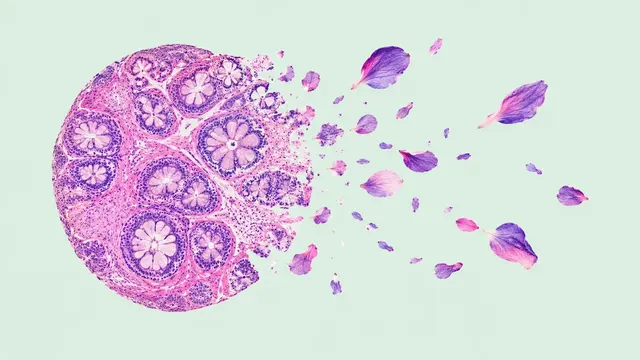
Colorectal Pathology reads the H&E slides of colorectal polypectomy specimens, biopsies and cancer resections, the highest-volume specimens in GI pathology. For a polyp, histology sets the colonoscopy surveillance interval; for a cancer, universal MSI or mismatch-repair testing is the guideline standard.
For polyps the run classifies tubular, tubulovillous and villous adenoma, sessile serrated lesion and hyperplastic polyp, and grades dysplasia. For carcinoma it returns the MSI-high/dMMR probability from H&E and a nine-class tissue map (tumour, stroma, lymphocytes, necrosis and debris, normal mucosa, muscle, adipose tissue, mucus, background) with the tumour–stroma ratio.
Decision support: a second look at the serrated-versus-hyperplastic distinction and at dysplasia grade, and an MSI probability that puts likely MSI-high cancers first in the queue for MMR immunohistochemistry or MSI PCR. Tangentially sectioned polyps and small biopsies are the hard cases.
GI and general surgical pathologists, residents, and screening programmes with large polyp volumes. Developers use the run for structured polyp data in a LIS or for optical-versus-histological diagnosis audits.
| Requirement | Why |
|---|---|
| H&E stain | All three sections read H&E morphology |
| Pixel spacing in µm/px | Tissue-class areas and tumour–stroma ratio are measured in mm² |
| Part label per polyp | Histology is reported per polyp, matching the colonoscopy site |
Polyp site, endoscopic findings or the clinical question can be passed in clinical_context.
| Section | Content |
|---|---|
| Polyp histology | Tubular, tubulovillous or villous adenoma, sessile serrated lesion or hyperplastic polyp, with dysplasia grade |
| MSI prediction | MSI-high/dMMR probability from H&E for carcinoma |
| Tissue map | Nine-class tissue segmentation with class areas and the tumour–stroma ratio |
| Draft report | Opt-in (options.report): an English narrative that interprets the result for the reader (findings, impression, limitations); every number is checked against the findings |
DiagnosticReport and IHE PaLM APSR for LIS export.The polyp-histology result pairs with the polyp characterisation of the Colonoscopy model (endo/colonoscopy), to
compare the optical diagnosis from the recorded colonoscopy with the final histology.
One run returns every section its input supports.
Prostate Biopsy Pathology
Prostate core biopsy read: cancer per core, Gleason and ISUP grade, % pattern 4, tumour length.
Any-Organ H&E Slide Analysis
Organ-agnostic H&E read: slide QC, tumour map, nuclei and mitoses, primary-site ranking, embeddings.
Breast Pathology
Breast case read from H&E, IHC and node slides: type, grade, biomarker scores, TILs and pN.
Lung Cancer Pathology
Lung biopsy and resection read: NSCLC subtype, IASLC grade, PD-L1 TPS and EGFR mutation probability.
Peripheral Blood Smear
Peripheral blood smear read: classified white cells, differential percentages and a blast alert.
Bone Marrow Aspirate
Marrow aspirate read: cell-by-cell myelogram, blast percentage, M:E ratio and dysplasia flags.