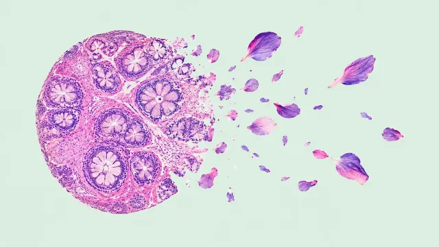
Prostate Biopsy Pathology
Prostate core biopsy read: cancer per core, Gleason and ISUP grade, % pattern 4, tumour length.
Pelvis4 sections
WSI· wsi
Organ-agnostic H&E read: slide QC, tumour map, nuclei and mitoses, primary-site ranking, embeddings.
This model is not available to run yet. Vote to show interest and get an email when it opens.
Any-Organ H&E Slide Analysis reads a single H&E whole-slide image from any organ. It is the general-purpose slide read in the library: use it when no organ-specific model fits, or when you need slide-level measurements rather than a diagnosis.
The run checks the scan first: tissue mask, artefacts (blur, folds, pen marks, bubbles, tears) and an adequacy verdict. It then maps tumour probability across the slide, divides the tissue into compartments (tumour, stroma, inflammation, necrosis, other), segments and classifies every nucleus, and finds mitotic figures with a count in the 2 mm² hot spot. For a metastasis or a cancer of unknown primary it ranks the likely tumour type and primary site. Tile and slide embeddings come with the result for developers.
Decision support and measurement. The result describes what is on the slide, and the primary-site ranking guides the choice of immunohistochemistry for a metastasis. Rare entities and lymphomas are the hard cases for an organ-agnostic tumour map.
Pathologists working up a metastasis of unknown origin, labs that want automatic scan QC before sign-out, and researchers and developers who need nuclei, mitosis and embedding outputs at slide scale through an API.
| Requirement | Why |
|---|---|
| H&E stain | IHC, cytology and haematology slides belong to other models |
| Pixel spacing in µm/px | Areas in mm², densities per mm² and the 2 mm² mitosis field depend on it |
| 20× or 40× scan | Nuclei and mitoses are read at native magnification |
| Imaged area and pixel matrix in the metadata | Needed to place maps and annotations on the pyramid |
The organ, specimen type and clinical question can go in clinical_context. A known organ narrows the
description; it does not turn the result into an organ-specific diagnosis.
| Section | Content |
|---|---|
| Slide quality | Tissue mask, artefact map and scan-adequacy verdict |
| Tumour detection | Tumour probability heatmap and tumour regions |
| Primary site | Ranked tumour types and likely primary sites for a metastasis or cancer of unknown primary |
| Tissue compartments | Map of tumour, stroma, inflammation, necrosis and other tissue, with area fractions |
| Nuclei | Instance masks classed as neoplastic, inflammatory, connective, dead (apoptotic or necrotic) or epithelial, with densities per mm² |
| Mitotic count | Typical and atypical mitotic figures and the count in the 2 mm² hot spot |
| Embeddings | Tile embeddings with coordinates and a slide embedding, as safetensors or HDF5 arrays |
| Draft report | Opt-in (options.report): an English narrative that interprets the result for the reader (findings, impression, limitations); every number is checked against the findings |
DiagnosticReport with Observation entries.The embedding arrays support slide search, cohort analytics and training your own slide classifiers without re-reading the image. Each array carries the tile coordinates and the encoder version it was computed with.
One run returns every section its input supports.
Prostate Biopsy Pathology
Prostate core biopsy read: cancer per core, Gleason and ISUP grade, % pattern 4, tumour length.
Breast Pathology
Breast case read from H&E, IHC and node slides: type, grade, biomarker scores, TILs and pN.
Colorectal Pathology
Colorectal polyp and cancer read: adenoma or serrated histology, dysplasia, MSI, tissue map.
Lung Cancer Pathology
Lung biopsy and resection read: NSCLC subtype, IASLC grade, PD-L1 TPS and EGFR mutation probability.
Peripheral Blood Smear
Peripheral blood smear read: classified white cells, differential percentages and a blast alert.
Bone Marrow Aspirate
Marrow aspirate read: cell-by-cell myelogram, blast percentage, M:E ratio and dysplasia flags.