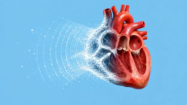
Echocardiography (TTE)
Full TTE read: views, LVEF, chambers, diastology, valves and effusion as ASE measurements.
Cardiac15 sections
US· cine
Per-zone lung ultrasound read: B-lines, sliding, consolidation, effusion, BLUE profile, LUS score.
This model is not available to run yet. Vote to show interest and get an email when it opens.

Lung Ultrasound reads the short clips a bedside physician records over the chest of a breathless or ventilated patient: one clip of three to six seconds per zone, at the BLUE points or over a 12-zone scheme, often from a handheld probe. Unlike most ultrasound, the lung exam reads artefacts rather than anatomy: A-lines, B-lines, the pleural line and its sliding.
The run returns a per-zone table (A pattern, B pattern, consolidation, effusion, sliding), combines the zones into a BLUE-protocol profile, and scores aeration zone by zone into a total LUS score. Together the sections address the time-critical questions: pneumothorax, cardiogenic pulmonary oedema or interstitial syndrome, pneumonia and pleural effusion.
Decision support for emergency and intensive-care physicians who scan and interpret their own lung clips, and a documented second read for the residents and nurses learning point-of-care lung ultrasound. Subcutaneous emphysema, bullae and pleurodesis can mimic pneumothorax, so the pneumothorax result is a probability backed by the sliding, lung-pulse and lung-point evidence.
Emergency physicians, intensivists, anaesthesiologists, pulmonologists, hospitalists and POCUS educators. Developers add the run to handheld-ultrasound apps, ICU charting and teleultrasound review.
| Requirement | Why |
|---|---|
| Cine, not stills | Sliding, lung pulse and B-line movement exist only over time; still images are refused for pneumothorax |
| 3 to 6 seconds per clip | At least one full respiratory cycle at the pleural line |
| Anterior zones on both sides | Pneumothorax is assessed on the anterior chest |
| Posterolateral (PLAPS) clips | Effusion and dependent consolidation are found there |
| 12 zones for the LUS score | The 0–36 total needs every zone scored |
The interpleural distance and the consolidation size need a calibrated or readable depth scale; without one, these sections return the pattern without a size.
The clinical question (dyspnoea, suspected pneumothorax, monitoring of aeration), ventilation status and age go in
clinical_context.
One JSON result in sections with a per-zone table, findings, measurements and scores, plus B-line masks and
overlay clips. A zone without a visible pleural line returns "insufficient view" rather than a pattern.
| Section | Content |
|---|---|
| B-lines | Presence and count per zone (0, 1–2, 3 or more, confluent), percentage of the intercostal space, location |
| Pneumothorax | Lung sliding present or absent, lung pulse, B-lines, lung point and a pneumothorax probability |
| Consolidation | Tissue-like pattern (hepatisation), shred sign, dynamic air bronchogram and size |
| Pleural effusion | Presence, simple or complex appearance, interpleural distance and a volume estimate |
| LUS aeration score | Aeration score 0–3 for each of 12 zones and the total from 0 to 36 |
| Draft report | Opt-in (options.report): an English narrative that interprets the result for the reader (findings, impression, limitations); every number is checked against the findings |
Each zone scores from 0 (normal aeration) to 3 (severe loss of aeration), and the 12 zone scores add up to the 0–36 total. B-line counts change with preset (harmonics, focus, depth) and probe, so repeat scores of a ventilated patient compare best on the same device and settings.
us/echo (focused section) and us/vascular (DVT) complete the breathless-patient work-up.us/fast uses the same pneumothorax read in trauma.One run returns every section its input supports.
Echocardiography (TTE)
Full TTE read: views, LVEF, chambers, diastology, valves and effusion as ASE measurements.
Obstetric Ultrasound
Trimester-aware pregnancy scan read: planes, biometry, GA, EFW, centiles, fluid and Doppler.
Thyroid and Neck Ultrasound
Thyroid nodule list with ACR TI-RADS level and FNA advice, gland volume and cervical lymph nodes.
Abdominal and Renal Ultrasound
Organ-by-organ abdominal read: liver, gallbladder, kidneys, spleen, aorta and paediatric bowel.
Breast Ultrasound
Breast lesion list with BI-RADS v2025 descriptors and final assessment, axillary nodes and ABUS.
eFAST Trauma Ultrasound
eFAST read per window: free fluid, pericardial effusion, pneumothorax and an overall result.