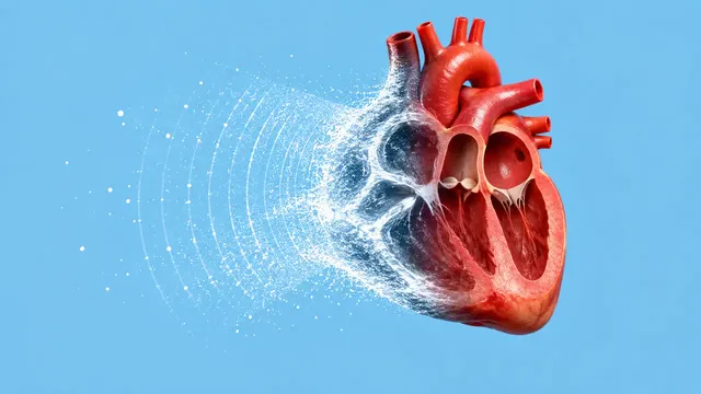
Echocardiography (TTE)
Full TTE read: views, LVEF, chambers, diastology, valves and effusion as ASE measurements.
Cardiac15 sections
US· cine
Region-aware MSK read: rotator cuff, carpal tunnel, synovitis, infant hip (Graf) and abscess.
This model is not available to run yet. Vote to show interest and get an email when it opens.

Musculoskeletal Ultrasound reads joint, tendon, nerve and soft-tissue studies, and the coronal hip images of infant hip screening. The run detects the body region from the images, so a shoulder study, a wrist, a set of finger joints, an infant hip or a soft-tissue swelling is each read with the questions that belong to it.
For the shoulder it grades the rotator cuff: partial- or full-thickness tears, tendinosis, bursitis and effusion. At the wrist it outlines the median nerve and measures its cross-sectional area at the tunnel inlet. In inflammatory arthritis it grades synovial hypertrophy and power Doppler per joint into the combined score. For the infant hip it places the Graf landmarks, measures the α and β angles and assigns a Graf type. For soft-tissue infections it separates abscess from cellulitis and sizes any drainable pocket.
Decision support in five different settings: the MSK radiologist or sports physician reporting a shoulder, the hand surgeon or neurophysiologist assessing carpal tunnel syndrome, the rheumatologist scoring synovitis at follow-up, the paediatric orthopaedist reading hip screening, and the emergency physician deciding whether an abscess can be drained. Anisotropy and manoeuvres not captured in stills limit what any reader can see, and the result says which views it relied on.
MSK radiologists, rheumatologists, orthopaedic and sports-medicine physicians, paediatric orthopaedists, hand surgeons, neurophysiologists and emergency physicians. Developers add the run to rheumatology registries, hip-screening programmes and orthopaedic reporting.
| Requirement | Why |
|---|---|
| Coronal hip image in the Graf standard plane | The α and β angles are valid only on the standard plane; other images are flagged |
| Transverse wrist image at the tunnel inlet | Median-nerve cross-sectional area is defined there |
| Grey-scale and power Doppler images of each joint | Synovial hypertrophy and Doppler signal are combined into the score |
SequenceOfUltrasoundRegions calibration |
Areas, thicknesses and cavity size in millimetres |
Age (in weeks for an infant hip), side, the joint or region examined and the clinical question go in
clinical_context.
One JSON result in sections; only the sections that match the detected region are filled, and each records the images it used. Findings carry the joint and side.
| Section | Content |
|---|---|
| Rotator cuff | Supraspinatus and other cuff tendons: partial- or full-thickness tear, tendinosis, bursitis, joint effusion |
| Carpal tunnel | Median-nerve outline, cross-sectional area at the tunnel inlet and a carpal tunnel syndrome probability |
| Synovitis | Grey-scale synovial hypertrophy and power-Doppler grade 0–3 per joint, with the combined score |
| Infant hip (Graf) | Ilium, acetabular roof and labrum landmarks; α and β angles; Graf type and femoral head coverage |
| Soft-tissue abscess | Abscess or cellulitis (cobblestoning), cavity size and whether a drainable pocket is present |
| Draft report | Opt-in (options.report): an English narrative that interprets the result for the reader (findings, impression, limitations); every number is checked against the findings |
The Graf type follows from the α and β angles measured on the image shown, and the angle lines are drawn on it, so the classification can be checked on the image. An α angle of 60° or more is a mature hip (type I). From 50° to 59° the hip is type II, split by age at 12 weeks into IIa and IIb. Below 50° the hip is critical or decentred (types IIc, D, III and IV), and the β angle and the position of the femoral head and labrum separate these types.
us/regional-anesthesia reads recorded nerve-block clips.us/vascular reads duplex studies of limb vessels.One run returns every section its input supports.
Echocardiography (TTE)
Full TTE read: views, LVEF, chambers, diastology, valves and effusion as ASE measurements.
Obstetric Ultrasound
Trimester-aware pregnancy scan read: planes, biometry, GA, EFW, centiles, fluid and Doppler.
Thyroid and Neck Ultrasound
Thyroid nodule list with ACR TI-RADS level and FNA advice, gland volume and cervical lymph nodes.
Abdominal and Renal Ultrasound
Organ-by-organ abdominal read: liver, gallbladder, kidneys, spleen, aorta and paediatric bowel.
Breast Ultrasound
Breast lesion list with BI-RADS v2025 descriptors and final assessment, axillary nodes and ABUS.
eFAST Trauma Ultrasound
eFAST read per window: free fluid, pericardial effusion, pneumothorax and an overall result.