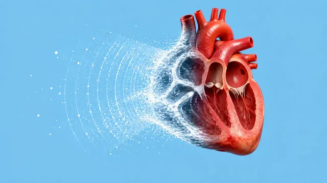
Echocardiography (TTE)
Full TTE read: views, LVEF, chambers, diastology, valves and effusion as ASE measurements.
Cardiac15 sections
US· cine
Preterm cranial ultrasound read: Papile IVH grade, ventricular index and dilatation flag.
This model is not available to run yet. Vote to show interest and get an email when it opens.

Neonatal Cranial Ultrasound reads the transfontanellar scans taken through the anterior fontanelle of preterm and sick newborns in the neonatal intensive care unit. Upload the coronal and sagittal (including parasagittal) stills, or the sweeps from which they come; the run sorts the images by plane and level before reading them.
It returns two sections. The haemorrhage section detects germinal-matrix and intraventricular haemorrhage and grades it with the Papile system from I to IV, identifying periventricular haemorrhagic infarction. The ventricular section measures the ventricular index, anterior-horn width and thalamo-occipital distance and flags post-haemorrhagic ventricular dilatation (PHVD), which neonatal teams follow closely after a bleed.
Decision support for the serial scans of a preterm infant: the first-week screening scan, the follow-up that decides whether a haemorrhage has extended, and the repeated measurements that track ventricular size after a bleed. Cranial ultrasound in the NICU is often performed at the cot side by neonatologists and read later by a radiologist; a structured grade and measurements make scans comparable from one week to the next.
Neonatologists, paediatric and neuro-radiologists, neonatal fellows and advanced neonatal nurse practitioners who scan, and paediatric neurosurgeons following ventricular dilatation. Developers add the run to NICU charting and neonatal follow-up registries.
| Requirement | Why |
|---|---|
| Coronal planes from frontal to occipital | Ventricular index and anterior-horn width are measured on coronal planes |
| Parasagittal planes through each lateral ventricle | Germinal-matrix haemorrhage, intraventricular blood and thalamo-occipital distance are seen there |
| Both hemispheres imaged | Each side is graded and measured separately |
SequenceOfUltrasoundRegions calibration |
Ventricular measurements in millimetres |
Gestational age at birth, age at the scan and the clinical question (screening, follow-up of a known haemorrhage,
growing head circumference) go in clinical_context.
One JSON result in sections with findings, measurements and classifications. Each grade and
measurement points to the image it was taken on, with calipers drawn on that image.
| Section | Content |
|---|---|
| Intraventricular haemorrhage | Germinal-matrix and intraventricular haemorrhage, Papile grade I–IV and periventricular haemorrhagic infarction |
| Ventricular size | Ventricular index, anterior-horn width and thalamo-occipital distance, with a PHVD flag |
| Draft report | Opt-in (options.report): an English narrative that interprets the result for the reader (findings, impression, limitations); every number is checked against the findings |
Papile grade I is confined to the germinal matrix. Grade II is blood in the ventricle without dilatation, and grade III distends the ventricle. Grade IV extends into the parenchyma; current practice describes this lesion as periventricular haemorrhagic infarction, and the result names it so. The ventricular index is compared with the Levene reference chart for postmenstrual age. Because the same three measurements are taken on every scan, serial scans line up and dilatation can be followed over weeks.
One run returns every section its input supports.
Echocardiography (TTE)
Full TTE read: views, LVEF, chambers, diastology, valves and effusion as ASE measurements.
Obstetric Ultrasound
Trimester-aware pregnancy scan read: planes, biometry, GA, EFW, centiles, fluid and Doppler.
Thyroid and Neck Ultrasound
Thyroid nodule list with ACR TI-RADS level and FNA advice, gland volume and cervical lymph nodes.
Abdominal and Renal Ultrasound
Organ-by-organ abdominal read: liver, gallbladder, kidneys, spleen, aorta and paediatric bowel.
Breast Ultrasound
Breast lesion list with BI-RADS v2025 descriptors and final assessment, axillary nodes and ABUS.
eFAST Trauma Ultrasound
eFAST read per window: free fluid, pericardial effusion, pneumothorax and an overall result.