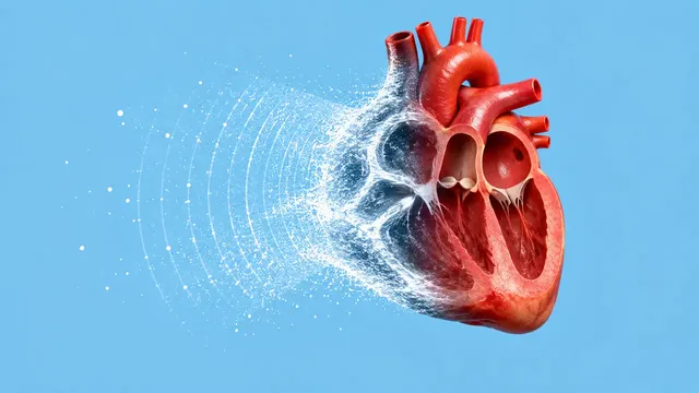
Echocardiography (TTE)
Full TTE read: views, LVEF, chambers, diastology, valves and effusion as ASE measurements.
Cardiac15 sections
US· cine
Ocular POCUS read: optic nerve sheath diameter and retinal detachment versus vitreous disease.
This model is not available to run yet. Vote to show interest and get an email when it opens.

Ocular Ultrasound (POCUS) reads the eye scans that emergency physicians record through the closed eyelid. Two questions bring a patient to this scan: sudden painless loss of vision, where the posterior segment must be checked for retinal detachment or vitreous haemorrhage, and suspected raised intracranial pressure, where the optic nerve sheath behind the globe is measured.
The run reads both eyes. For the posterior segment it uses the clips taken while the patient moves the eye, because the way a membrane or an opacity moves separates the three main causes of a mobile echo in the vitreous: retinal detachment, posterior vitreous detachment and vitreous haemorrhage. For the optic nerve it measures the sheath diameter 3 mm behind the globe in two planes per eye and raises a flag when the measurement suggests raised intracranial pressure.
Decision support for the emergency or critical-care physician deciding how urgently to involve ophthalmology or neurosurgery, and a structured record of a bedside scan that is otherwise rarely documented. The result shows the frames and calipers it relied on so the reader can check them.
Emergency physicians, intensivists, neurocritical-care teams, ophthalmologists receiving referrals, and POCUS educators teaching ocular scanning. Developers add the run to handheld-ultrasound apps and emergency documentation.
| Requirement | Why |
|---|---|
| Both eyes scanned | Each eye is read and reported separately |
| Transverse and sagittal plane of each optic nerve | The sheath diameter is measured in two planes per eye |
| Optic nerve shown in its long axis behind the globe | The measurement point lies 3 mm behind the globe on that axis |
| Eye-movement clip for the posterior segment | Detachment and vitreous disease are distinguished by how they move |
Calibrated depth (SequenceOfUltrasoundRegions or a readable depth scale) |
The sheath diameter is a millimetre value; without a scale, only the posterior-segment read is returned |
Handheld clips are common for this scan. The result names the source of the scale used for every diameter.
Age and the clinical question (sudden vision loss, head injury, suspected raised intracranial pressure) go in
clinical_context.
One JSON result in two sections with findings and measurements for each eye. The sheath diameter carries both
plane values and the frames they were taken on, with calipers drawn as overlays.
| Section | Content |
|---|---|
| Optic nerve sheath diameter | ONSD 3 mm behind the globe in two planes per eye, with a raised-ICP flag |
| Retinal detachment | Retinal detachment, posterior vitreous detachment or vitreous haemorrhage, with the supporting eye-movement frames |
| Draft report | Opt-in (options.report): an English narrative that interprets the result for the reader (findings, impression, limitations); every number is checked against the findings |
Optic nerve sheath diameter is a non-invasive sign of raised intracranial pressure, not a pressure value; the flag prompts further assessment. A posterior-segment finding is reported per eye with the clip that shows it moving.
us/fast and us/lung read the other emergency point-of-care protocols.One run returns every section its input supports.
Echocardiography (TTE)
Full TTE read: views, LVEF, chambers, diastology, valves and effusion as ASE measurements.
Obstetric Ultrasound
Trimester-aware pregnancy scan read: planes, biometry, GA, EFW, centiles, fluid and Doppler.
Thyroid and Neck Ultrasound
Thyroid nodule list with ACR TI-RADS level and FNA advice, gland volume and cervical lymph nodes.
Abdominal and Renal Ultrasound
Organ-by-organ abdominal read: liver, gallbladder, kidneys, spleen, aorta and paediatric bowel.
Breast Ultrasound
Breast lesion list with BI-RADS v2025 descriptors and final assessment, axillary nodes and ABUS.
eFAST Trauma Ultrasound
eFAST read per window: free fluid, pericardial effusion, pneumothorax and an overall result.