Echocardiography (TTE)
Full TTE read: views, LVEF, chambers, diastology, valves and effusion as ASE measurements.
Cardiac15 sections
US· cine
Recorded nerve-block clip read: nerves, vessels, fascia, pleura and needle tip for QA and teaching.
This model is not available to run yet. Vote to show interest and get an email when it opens.
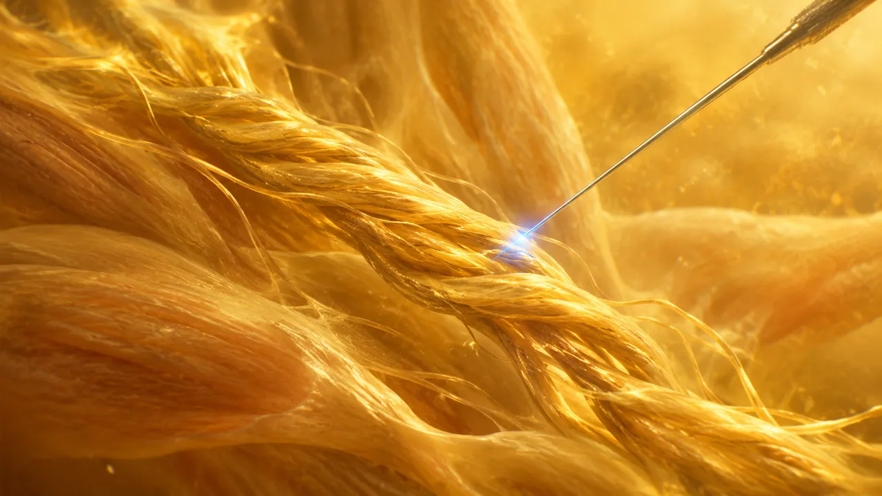
Nerve Block Ultrasound reads a recorded clip of an ultrasound-guided regional anaesthesia procedure. Upload one block clip and name the block; the run identifies the structures the anaesthesiologist aims for and avoids, and follows the needle through the recording.
It returns two sections. The first outlines, on every frame, the nerves or plexus targeted by that block and the landmarks around them: arteries, fascial planes and the pleura. The second locates the needle shaft and tip frame by frame and reconstructs its trajectory. Together they answer, after the procedure, whether the target plane was adequate and where the needle went during the procedure.
The run reads clips after they were recorded. Its uses are training, quality assurance and documentation: a resident's block reviewed with the target and needle path marked, a department auditing its block recordings, and a structured note of the sono-anatomy seen.
Anaesthesiologists and anaesthesia residents, regional-anaesthesia fellowship programmes, simulation and teaching centres, emergency physicians who perform blocks, and quality teams. Developers add the run to training platforms, procedure logs and departmental QA tools.
| Requirement | Why |
|---|---|
| Cine, not a still | The needle is followed across frames and its trajectory is built from the sequence |
| Block type given | The target structures and their expected neighbours depend on the block |
| Target region in view | Plane adequacy is judged on the frames where the target is visible |
| Needle visible in the clip | Tip and shaft can only be located on frames where the needle appears |
Screen recordings from a handheld device or a teaching station are accepted, since this read does not depend on millimetre calibration.
The purpose of the review (teaching, complication review, documentation) can go in clinical_context.
One JSON result in two sections. Masks are returned per frame as DICOM SEG, and an overlay video shows the structures and the needle on the original clip.
| Section | Content |
|---|---|
| Nerves and landmarks | Per-frame segmentation of the target nerve or plexus and surrounding landmarks (arteries, fascia, pleura), with target-plane adequacy |
| Needle tip and trajectory | Needle shaft and tip location per frame and the reconstructed trajectory |
| Draft report | Opt-in (options.report): an English narrative that interprets the result for the reader (findings, impression, limitations); every number is checked against the findings |
Target-plane adequacy reflects whether the expected structures for the named block were visible together. The needle section reports where the tip was seen; frames without a visible tip are marked as such.
us/msk reads nerve, tendon and joint studies, including the median nerve at the carpal tunnel.One run returns every section its input supports.
Echocardiography (TTE)
Full TTE read: views, LVEF, chambers, diastology, valves and effusion as ASE measurements.
Obstetric Ultrasound
Trimester-aware pregnancy scan read: planes, biometry, GA, EFW, centiles, fluid and Doppler.
Thyroid and Neck Ultrasound
Thyroid nodule list with ACR TI-RADS level and FNA advice, gland volume and cervical lymph nodes.
Abdominal and Renal Ultrasound
Organ-by-organ abdominal read: liver, gallbladder, kidneys, spleen, aorta and paediatric bowel.
Breast Ultrasound
Breast lesion list with BI-RADS v2025 descriptors and final assessment, axillary nodes and ABUS.
eFAST Trauma Ultrasound
eFAST read per window: free fluid, pericardial effusion, pneumothorax and an overall result.