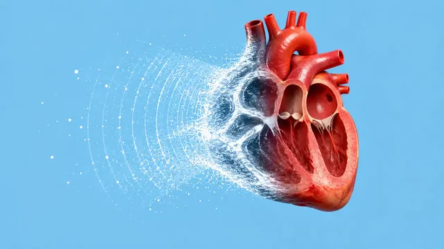
Echocardiography (TTE)
Full TTE read: views, LVEF, chambers, diastology, valves and effusion as ASE measurements.
Cardiac15 sections
US· cine
Duplex read: carotid stenosis and IMT, DVT compression and peripheral arterial waveforms.
This model is not available to run yet. Vote to show interest and get an email when it opens.
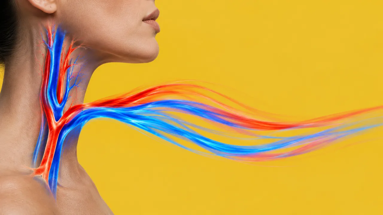
Vascular Duplex Ultrasound reads vascular-lab and bedside studies that combine B-mode, colour and spectral Doppler: carotid duplex after a TIA or stroke, lower-limb venous compression for deep vein thrombosis, including the two- or three-point scans done by emergency physicians, and arterial duplex for peripheral arterial disease.
For each vessel the run returns its findings. Carotid velocities are read from the spectral strips, from the sonographer's on-screen values or from vendor SR, and combined with plaque into a stenosis category; the common carotid far wall is measured for intima-media thickness; compression clips are read segment by segment for compressibility and thrombus; arterial waveforms are classified and stenoses graded by velocity ratio.
Decision support for the vascular sonographer, the vascular physician who signs the study, and the emergency doctor deciding whether a leg is positive for DVT. Angle correction and lab-specific velocity criteria change a stenosis grade, so the result names the criteria it applied. Compression clips that do not show full probe pressure are flagged.
Vascular surgeons, angiologists and vascular-medicine physicians, neurologists in stroke units, vascular sonographers, radiologists and emergency physicians. Developers add the run to vascular-lab reporting and stroke or venous-thromboembolism pathways.
| Requirement | Why |
|---|---|
| Spectral strips with a calibrated velocity axis | PSV, EDV and ratios are read from the spectrum |
| Angle-corrected ICA and CCA samples | Velocity criteria assume a correct insonation angle |
| Longitudinal CCA B-mode with a clear far wall | IMT is measured on the far wall |
| Cine showing full compression of each venous segment | Compressibility is a motion finding; stills are refused for it |
SequenceOfUltrasoundRegions calibration |
IMT and plaque in millimetres |
The clinical question (TIA or stroke, suspected DVT, claudication) and the side examined go in clinical_context.
One JSON result in sections with findings, measurements and classifications per vessel and side. Each velocity
keeps its provenance: traced by the model, read from the screen or taken from vendor SR.
| Section | Content |
|---|---|
| Carotid stenosis | ICA PSV and EDV, ICA/CCA ratio and plaque, with the stenosis category per side |
| Carotid intima-media thickness | Far-wall IMT of the common carotid (mean and maximum) and plaque presence |
| Deep vein thrombosis | Compressibility of each venous segment, intraluminal thrombus and a positive or negative DVT result |
| Peripheral arterial duplex | Waveform class (multiphasic to monophasic) and stenosis by PSV ratio |
| Draft report | Opt-in (options.report): an English narrative that interprets the result for the reader (findings, impression, limitations); every number is checked against the findings |
Carotid stenosis grading is a deterministic rule applied to the velocities shown in the result, so each category can be checked against its numbers. Under the SRU 2003 criteria, an ICA PSV of 125–230 cm/s with visible plaque indicates 50–69 % stenosis, and a PSV above 230 cm/s indicates 70 % or more; the ICA/CCA ratio and EDV support the category. A DVT result applies only to the segments imaged; segments not shown are listed.
us/echo and us/echo-tee complete a cardio-embolic stroke work-up.us/lung and us/echo join the DVT read in the breathless patient.One run returns every section its input supports.
Echocardiography (TTE)
Full TTE read: views, LVEF, chambers, diastology, valves and effusion as ASE measurements.
Obstetric Ultrasound
Trimester-aware pregnancy scan read: planes, biometry, GA, EFW, centiles, fluid and Doppler.
Thyroid and Neck Ultrasound
Thyroid nodule list with ACR TI-RADS level and FNA advice, gland volume and cervical lymph nodes.
Abdominal and Renal Ultrasound
Organ-by-organ abdominal read: liver, gallbladder, kidneys, spleen, aorta and paediatric bowel.
Breast Ultrasound
Breast lesion list with BI-RADS v2025 descriptors and final assessment, axillary nodes and ABUS.
eFAST Trauma Ultrasound
eFAST read per window: free fluid, pericardial effusion, pneumothorax and an overall result.