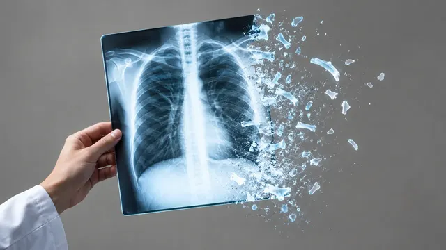
Chest X-ray
Chest radiograph read: urgent flags, findings with boxes, device tips, CTR and interval change.
Chest17 sections
XR· image-2d
DXA read: T-scores by ISCD rules, VFA fractures, aortic calcification and body composition.
This model is not available to run yet. Vote to show interest and get an email when it opens.
DXA reads a dual-energy X-ray absorptiometry study: lumbar spine, hip and forearm scans, with a lateral-spine vertebral fracture assessment (VFA) image and a whole-body scan when they were acquired. Bone mineral density is measured by the scanner's own software, so the run takes the BMD values from the vendor result pages instead of measuring them again, and applies the ISCD reporting rules to them: T- and Z-scores per site, the WHO category, the change against the prior scan judged by least-significant change, and a FRAX prompt.
Where the images allow more, the run reads them too. On the VFA image it grades each vertebra for fracture and scores calcification of the abdominal aorta; on a whole-body scan it returns regional lean and fat mass. A quality check flags positioning and region-of-interest errors and the vertebrae that should be excluded from the spine result.
Decision support for the clinician who reports or acts on the scan. Degenerative change and vertebral fractures can falsely raise spine BMD, and a vertebral fracture changes fracture risk and treatment whatever the T-score; both are easy to overlook on a printed result page. The run brings density, fractures and scan quality into one structured result.
GPs, endocrinologists, rheumatologists, geriatricians, radiologists and technologists in bone-density services. Developers can turn DXA exports into structured data for an osteoporosis registry or fracture liaison service.
| Requirement | Why |
|---|---|
| Vendor BMD results (DICOM SR or result pages) | BMD values are parsed from them, not re-measured |
| Scan images with the vendor regions of interest | Positioning, ROI placement and excluded vertebrae are checked on the images |
| Age and sex | WHO T-score categories apply to postmenopausal women and men aged 50 and over; younger adults are reported by Z-score |
| Lateral-spine VFA image | Needed for vertebral fracture grades and the aortic calcification score |
| Whole-body scan | Needed for body composition |
| Prior study from the same scanner | Change is judged against the least-significant change |
Menopausal status, risk factors for FRAX (previous fracture, glucocorticoid use, parental hip fracture, smoking) and
current treatment in clinical_context complete the fracture-risk prompt.
One JSON result in sections, with every score traced to the vendor value or image it came from.
| Section | Content |
|---|---|
| Bone density | Vendor BMD results per site parsed into T- and Z-scores, the WHO category by ISCD rules, change versus the prior against the least-significant change, and a FRAX prompt |
| Scan quality | Positioning, ROI placement, artefacts and vertebrae to exclude from the spine result |
| Vertebral fracture assessment | Genant grade for each vertebra on the VFA image |
| Aortic calcification | Abdominal aortic calcification score on the VFA image |
| Body composition | Regional lean and fat mass, appendicular lean mass index (ALMI), visceral adipose tissue and a sarcopenia flag |
| Draft report | Opt-in (options.report): an English narrative that interprets the result for the reader (findings, impression, limitations); every number is checked against the findings |
One run returns every section its input supports.
Chest X-ray
Chest radiograph read: urgent flags, findings with boxes, device tips, CTR and interval change.
Musculoskeletal X-ray
Bone and joint radiograph read: fractures, dislocations, bone age, osteoarthritis and alignment.
Abdominal X-ray
Abdominal film read: bowel gas pattern, free air, radio-opaque stones and swallowed objects.
NICU Radiograph
Neonatal babygram read: umbilical line and tube tips by vertebral level, plus NEC signs.
Spine X-ray
Spine radiograph read: scoliosis curves, vertebral fractures, sagittal balance and slip grades.
Hysterosalpingogram
HSG read: fill and spill for each fallopian tube, with occlusion level and hydrosalpinx.