Chest X-ray
Chest radiograph read: urgent flags, findings with boxes, device tips, CTR and interval change.
Chest17 sections
XR· image-2d
Neonatal babygram read: umbilical line and tube tips by vertebral level, plus NEC signs.
This model is not available to run yet. Vote to show interest and get an email when it opens.
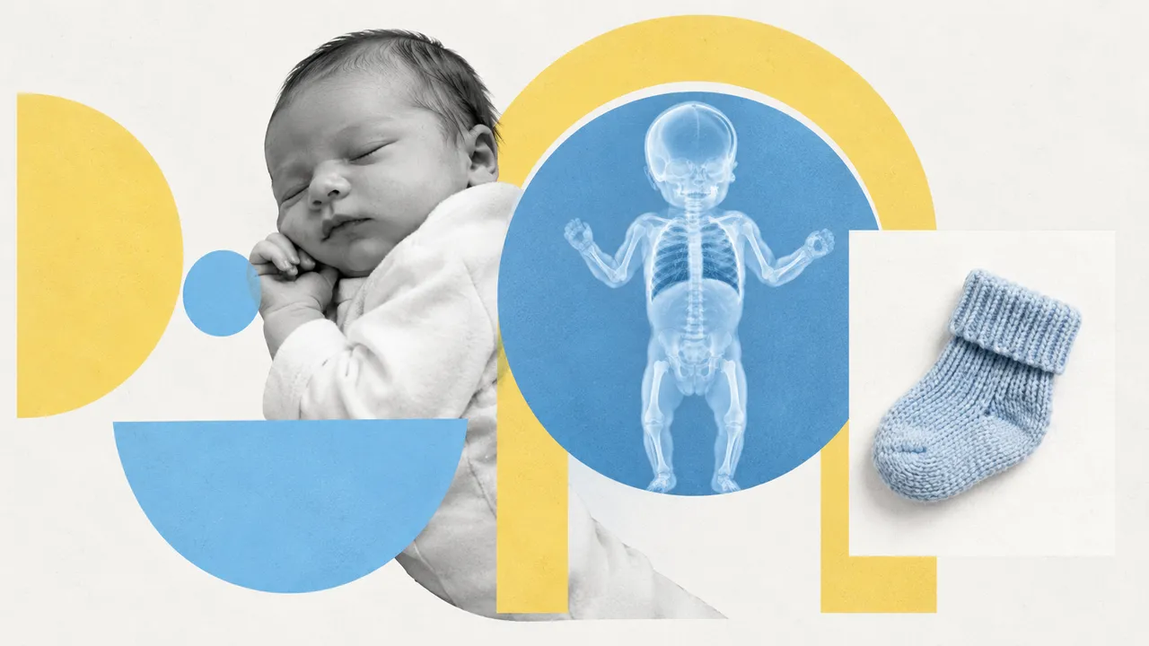
NICU Radiograph reads the combined chest and abdomen film of a newborn, the "babygram" taken after every umbilical line or tube placement and repeatedly in ventilated infants. It is a separate model from the adult chest film because the image (supine AP, chest and abdomen together, very small anatomy), the devices and the findings are different.
The run finds each endotracheal tube, umbilical venous catheter (UVC), umbilical arterial catheter (UAC), nasogastric or orogastric tube and PICC, locates its tip by vertebral level and flags a malposition. On the abdomen it looks for the signs of necrotising enterocolitis: pneumatosis intestinalis, portal venous gas, pneumoperitoneum and a fixed loop, and suggests the radiographic part of a modified Bell stage; clinical signs complete the staging.
Decision support for the first reader of the film, often a neonatology resident at night, who must decide whether a freshly placed line can be used or needs to be repositioned. Line malposition is frequent and time-critical; the tip level and the target zone are shown on the image. NEC signs come with the modified Bell stage they suggest.
Neonatologists, neonatal nurse practitioners, paediatric residents, paediatric surgeons and paediatric radiologists. Developers can connect the run to a NICU imaging workflow so every line-check film gets a structured tip report.
| Requirement | Why |
|---|---|
| Chest and upper abdomen in the field | UVC and UAC tips are judged against the vertebral bodies and the diaphragm |
| The whole abdomen for NEC | Pneumatosis, portal venous gas and fixed loops can be anywhere in the bowel |
| Patient age (days) or birth date | Selects the neonatal path; older children go to Chest X-ray |
| Low rotation | Rotation shifts the apparent tip level against the spine |
The thymus shadow, skin folds and overlapping monitoring leads are the known mimics on neonatal films.
Gestational age and the device just placed in clinical_context; a prior film in
prior_files shows device moves and the evolution of bowel signs.
A JSON result with a device list (type, tip coordinates, tip vertebral level, status) and a NEC section.
| Section | Content |
|---|---|
| Lines and tubes | ETT, UVC, UAC, NG/OG tube and PICC: tip vertebral level, position against the target and a malposition flag |
| Necrotising enterocolitis | Pneumatosis intestinalis, portal venous gas, pneumoperitoneum, fixed loop, and the modified Bell stage these signs suggest |
| Draft report | Opt-in (options.report): an English narrative that interprets the result for the reader (findings, impression, limitations); every number is checked against the findings |
| Device | Target used for the flag |
|---|---|
| UAC (high position) | Tip between T6 and T9 |
| UVC | Tip at the junction of the inferior vena cava and the right atrium |
| ETT | Tip in the mid-trachea |
Each device is reported as normal, borderline or abnormal, with its distance to the target landmark when the film is calibrated.
One run returns every section its input supports.
Chest X-ray
Chest radiograph read: urgent flags, findings with boxes, device tips, CTR and interval change.
Musculoskeletal X-ray
Bone and joint radiograph read: fractures, dislocations, bone age, osteoarthritis and alignment.
Abdominal X-ray
Abdominal film read: bowel gas pattern, free air, radio-opaque stones and swallowed objects.
Spine X-ray
Spine radiograph read: scoliosis curves, vertebral fractures, sagittal balance and slip grades.
DXA (Bone Densitometry)
DXA read: T-scores by ISCD rules, VFA fractures, aortic calcification and body composition.
Hysterosalpingogram
HSG read: fill and spill for each fallopian tube, with occlusion level and hydrosalpinx.