Chest X-ray
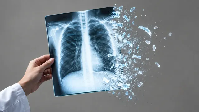
Chest radiograph read: urgent flags, findings with boxes, device tips, CTR and interval change.
Chest17 sections
XR· image-2d
HSG read: fill and spill for each fallopian tube, with occlusion level and hydrosalpinx.
This model is not available to run yet. Vote to show interest and get an email when it opens.

Hysterosalpingogram reads the fluoroscopy images of an HSG, the X-ray test of the uterine cavity and the fallopian tubes in a fertility work-up. Contrast is injected through the cervix while a short series of images is taken; the run follows it from the cavity into each tube and out into the peritoneal cavity.
For each tube it answers the two questions of the test: does contrast fill the tube, and does it spill freely from the fimbrial end? The answer per side is one of four: patent, proximal occlusion, distal occlusion, or hydrosalpinx (a distended, distally blocked tube). The images that show fill and spill are returned with the answer.
Decision support for the clinician who performs and reports the HSG, very often a gynaecologist or fertility specialist rather than a radiologist. A structured per-tube answer, tied to the frames that show it, gives every operator's report the same structure and supports the next step in the fertility plan.
Gynaecologists, reproductive endocrinologists and fertility specialists, radiologists who perform HSG, and fertility clinic teams. Developers can connect the run to a fertility clinic record or a fluoroscopy archive to store tubal findings as structured data.
| Requirement | Why |
|---|---|
| Images from early filling to late spill | Patency is judged from the sequence, not one frame |
| Both cornual regions and adnexa in the field | Each tube is assessed from its origin to its fimbrial end |
| Image orientation or laterality marker | The answer is given per side |
| A late image after injection | Shows whether the spilled contrast spreads freely in the pelvis |
Age, duration of infertility, previous pelvic infection or surgery and a previous HSG can be given in
clinical_context; they shape the guidance in the result.
One JSON result whose patency section holds a per-tube classification, with key images marking fill and spill.
| Section | Content |
|---|---|
| Tubal patency | Per tube: patent, proximal occlusion, distal occlusion or hydrosalpinx, with the key images |
| Draft report | Opt-in (options.report): an English narrative that interprets the result for the reader (findings, impression, limitations); every number is checked against the findings |
| Answer | Image pattern |
|---|---|
| Patent | The tube fills along its length and contrast spills freely into the peritoneal cavity |
| Proximal occlusion | No contrast enters the tube beyond the uterine cornu |
| Distal occlusion | The tube fills but contrast does not spill from the fimbrial end |
| Hydrosalpinx | The tube is distended with contrast and blocked at its distal end |
One run returns every section its input supports.
Chest X-ray
Chest radiograph read: urgent flags, findings with boxes, device tips, CTR and interval change.
Musculoskeletal X-ray
Bone and joint radiograph read: fractures, dislocations, bone age, osteoarthritis and alignment.
Abdominal X-ray
Abdominal film read: bowel gas pattern, free air, radio-opaque stones and swallowed objects.
NICU Radiograph
Neonatal babygram read: umbilical line and tube tips by vertebral level, plus NEC signs.
Spine X-ray
Spine radiograph read: scoliosis curves, vertebral fractures, sagittal balance and slip grades.
DXA (Bone Densitometry)
DXA read: T-scores by ISCD rules, VFA fractures, aortic calcification and body composition.