Chest X-ray
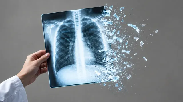
Chest radiograph read: urgent flags, findings with boxes, device tips, CTR and interval change.
Chest17 sections
XR· image-2d
Spine radiograph read: scoliosis curves, vertebral fractures, sagittal balance and slip grades.
This model is not available to run yet. Vote to show interest and get an email when it opens.
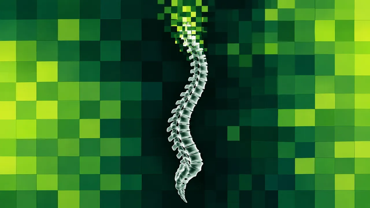
Spine X-ray reads cervical, thoracic, lumbar and standing whole-spine radiographs, frontal and lateral. The run locates each vertebra, then reports coronal alignment, vertebral body height, sagittal balance and slip between levels, with the landmarks it measured from drawn on the image.
Two problems drive it. Osteoporotic vertebral fractures are under-reported on routine spine and chest films, and they change treatment. Manual Cobb and sagittal measurements are slow and vary by about five degrees between readers; the run measures each curve and parameter from landmarks drawn on the image, the same way on every follow-up film.
Decision support for the reader of a spine film: a scoliosis or deformity measurement for the spine surgeon, a fracture check for the GP or radiologist reading a lateral thoracolumbar view, and a slip grade for back-pain work-up. Every measurement carries the landmarks it was measured from.
Radiologists, orthopaedic and spine surgeons, paediatric scoliosis clinics, osteoporosis services and GPs. Developers can add automated Cobb angles or vertebral fracture screening to a PACS or surgical-planning workflow through the API.
| Requirement | Why |
|---|---|
| Standing AP whole spine | Cobb angles, curve apex and Lenke curve pattern |
| Standing lateral whole spine including the pelvis | SVA, pelvic incidence, pelvic tilt and sacral slope need the femoral heads and the S1 endplate |
| Lateral thoracic or lumbar view | Vertebral height loss is graded from T4 to L4 on lateral projections |
| Pixel spacing with magnification | SVA and slip distances in millimetres; angles do not depend on it |
Stitching artefacts, rotation, transitional vertebrae (which affect level counting) and implants are flagged where they limit a measurement.
Age and the clinical question (adolescent idiopathic scoliosis, adult deformity, suspected osteoporotic fracture, back
pain) in clinical_context; a prior film in prior_files for curve progression.
A sectioned JSON result with vertebral landmarks, measurements and grades; overlays show the endplate lines and angles.
| Section | Content |
|---|---|
| Cobb angle | Vertebral landmarks, proximal thoracic, main thoracic and thoracolumbar/lumbar Cobb angles, curve apex and Lenke curve type |
| Vertebral fractures | Per-vertebra height loss and Genant grade 0–3 from T4 to L4 |
| Sagittal alignment | SVA, PI, PT, SS, lumbar lordosis, thoracic kyphosis and PI–LL mismatch |
| Spondylolisthesis | Slip percentage and Meyerding grade per level, and dynamic instability on flexion–extension films |
| Draft report | Opt-in (options.report): an English narrative that interprets the result for the reader (findings, impression, limitations); every number is checked against the findings |
One run returns every section its input supports.
Chest X-ray
Chest radiograph read: urgent flags, findings with boxes, device tips, CTR and interval change.
Musculoskeletal X-ray
Bone and joint radiograph read: fractures, dislocations, bone age, osteoarthritis and alignment.
Abdominal X-ray
Abdominal film read: bowel gas pattern, free air, radio-opaque stones and swallowed objects.
NICU Radiograph
Neonatal babygram read: umbilical line and tube tips by vertebral level, plus NEC signs.
DXA (Bone Densitometry)
DXA read: T-scores by ISCD rules, VFA fractures, aortic calcification and body composition.
Hysterosalpingogram
HSG read: fill and spill for each fallopian tube, with occlusion level and hydrosalpinx.